



All In Nutritionals
Home Blog Page 362

When patients want to get off their psychotropic medication(s)

0

When patients want to get off their psychotropic medication(s)

Effective strategies for limiting withdrawal and destabilization

For over a decade I have focussed the majority of my clinical practice on the evaluation and treatment of mental health disorders. One of the most common requests I receive from patients is their desire to reduce or discontinue their psychotropic medications. It is entirely appropriate for patients to request this. eir psychotropic drugs might be doing more harm than good and creating many objectionable side e ects, which are limiting rather than enhancing quality of life (Moncrie 2006a). ey might want to see how they feel and function once their psychotropic drugs are reduced and/or discontinued. ey might also wish to be drug-free. When this request it made, I usually tell then something along the lines of:

“I need to see that you are stable for at least six-months before there is any consideration for reducing and/or discontinuing your psychotropic medication. By stability I mean: getting along well with family and friends; maintaining some type of regular work or employment, such as school, a volunteer position, or a career; and having no more disabling symptoms of your psychiatric disorder.”

This is usually well received since most patients prefer to work in a slow, methodical fashion, rather than becoming destabilized should this process be rushed.

Difficulties in Overcoming Pharmacological Dependence

Pharmacological dependence is an expected and biological adaptation of the body becoming long habituated to the presence of psychotropic drugs (O’Brien 2005). Since psychotropic drugs can induce unpredictable global reactions when used properly (Moncrieff 2006b, Moncrieff 2009), there are no reliable ways to predict how patients will overcome pharmacological dependence once their psychotropic drugs are tapered and eventually withdrawn. Every patient’s withdrawal process is unique as is their susceptibility to develop withdrawal side effects (Read 2005).

Discontinuation Reactions Following Cessation of Psychotropic Drugs

Some patients do fine and experience little withdrawal, while other patients have become so habituated and dependent on their psychotropic drugs that stopping them is paramount to disaster. While no ethical clinician would tell a patient to abruptly stop his/ her psychotropic drugs, it is important for clinicians to recognize discontinuation reactions from the common classes of psychotropic drugs (Table 1).
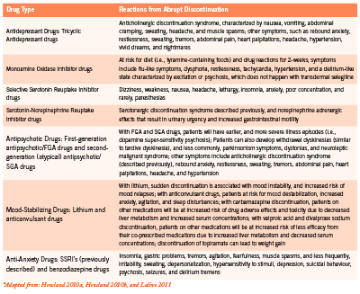
Table 1. Possible Discontinuation Reactions when Psychotropic Drugs are Stopped Abruptly*

Tapering Patients off their Psychotropic Drugs

When it is appropriate for this to happen, I usually communicate in writing to the patient’s prescribing clinician (i.e., the general/ family practitioner or psychiatrist) that the patient is doing well and wants to try discontinuing one of his/her psychotropic drugs. If the prescribing clinician does not agree to any plan of psychotropic drug tapering, despite the patient’s wishes and noted stability, it is entirely appropriate for the patient and non-prescribing clinician to proceed with a tapering plan. In such a circumstance, it might be suitable to draft a waiver outlining all potential risks and benefits of psychotropic drug tapering with the patient. The waiver would have to clearly delineate that this is what the patient wants, and that the patient is willing to assume all risks (e.g., destabilization and hospitalization) associated with the tapering process.

Screen Shot 2014-07-06 at 12.53.48 PM

Since there is a scarcity of clinical research pertaining to tapering patients off their psychotropic drugs, there are no clearly defined standards informing clinicians about the best and most appropriate method of tapering. Patients are just as likely to succeed in coming off their psychotropic drugs whether or not they have the support and endorsement from their prescribing clinicians (Darton 2005). The published data from one organization found that 53% of patients were successful in coming off their psychotropic drugs – even though this was either against their doctors’ advice or occurred without their doctors’ knowledge – compared to 44% that had their doctors’ approval (Read 2005).

The tapering plan should involve one drug at a time and should involve the drug with the longest elimination half-life first. Psychotropic drugs with longer elimination half-lives (i.e., more than 24 hours) are easier to taper from since their withdrawal reactions tend to be less severe than drugs with shorter elimination half-lives (i.e., 24 hours or less). Psychotropic drugs with short elimination half-lives can produce significant and quick withdrawal reactions. Considerations should be made to switch patients from psychotropic drugs with short elimination half-lives to drugs with longer elimination half-lives prior to tapering, as this will increase the chances of a positive experience and successful outcome. A psychotropic drug like vanlafaxine (has an elimination half-life of around five hours) can cause significant withdrawal and possible destabilization. A psychotropic drug like fluoxetine, on the other hand, can be used as a substitute for vanlafaxine, allowing for a more successful tapering plan since the elimination half-life of this drug is four to six days (Darton 2005).

Overall, the tapering plan needs to be very slow and gradual, which reduces the potential for relapse (Moncrieff 2006a). The patient should never be made to feel like a failure during this process since coming off of psychotropic drugs is very difficult due to the “physiological and psychological manifestations of the biological effects caused by the withdrawal of a regularly administered drug” (Moncrieff 2006a). For patients to successfully come of their psychotropic drugs they need to: (1) be well prepared and informed of the process; (2) have a positive attitude; (3) be mindful that they will experience strong emotions throughout tapering and possibly for months after; and (4) receive some type of regular psychological support (e.g., psychotherapy, art therapy, support groups, etc) during the tapering and perhaps beyond (Darton 2005, Hall 2007).

A good rule of thumb is for the tapering duration to be for the same length of time of previous psychotropic use (Hall 2007). For example, if a patient was on a psychotropic drug for six months, the tapering process should be six months. The only exception involves patients that have been on psychotropic drugs for five or more years. The tapering process for these patients should be approximately 18-24 months (Hall 2007).

One report suggests that for each unspecified tapering period, there should be a 10% reduction in dose, and that the psychotropic drug is reduced by subsequent 10% reductions until the patient is comfortably off his/her medication (Darton 2005). Another report suggests that the psychotropic drug is tapered down every two weeks, and that with each two-week tapering period, the dose is reduced by 10% (Hall 2007). If a patient, for example, begins the tapering process on a drug that is 400 mg once daily, than the dose to take for two weeks would be 360 mg. After two weeks, the dose would be further reduced by another 10% to 320 mg and so on. This process would continue until the psychotropic drug is fully discontinued. If, at any time during the tapering, the patient feels that the reduction is too marked or there are troublesome withdrawal reactions such as anxiety and sleep disturbances, the patient can remain on the previous tapered dose until he/she feels well enough to proceed.

Improving the Odds of a Successful Outcome

It is not uncommon for patients to experience problems even from small reductions in their psychotropic drugs. This is usually misattributed to a re-emergence of their underlying psychiatric disorder as opposed to problems related to psychotropic drug withdrawal (Moncrieff 2006a). When this happens, it is common for such patients to be shamed by family members, psychiatrists, and support workers into the notion that they cannot maintain their psychological health without depending on psychotropic drugs for the rest of their lives, thus forcing patients back onto long-term treatment (Moncrieff 2006a). Darton (2005) reported that patients with mental health issues are too often denied the chance to come off their medications, to take risks, and potentially make their own mistakes. If, on the other hand, family members are helpful, supportive, and encouraging, this will reduce risks associated from tapering off psychotropic drugs (Darton 2005).

chance to come off their medications, to take risks, and potentially make their own mistakes. If, on the other hand, family members are helpful, supportive, and encouraging, this will reduce risks associated from tapering off psychotropic drugs (Darton 2005).

Supporting the Tapering Process with Specific Natural Health Products

There are specific natural health products that I have found helpful when used during and after the tapering process to mitigate withdrawal, and prevent potential relapses, and possibly the need to resume previous psychotropic drugs. Melatonin is being formally studied to ascertain its benefits among schizophrenic patients withdrawing from long-term benzodiazepine use (Baandrup 2011). Previous reports have shown some efficacy in reducing benzodiazepine-withdrawal-associated sleep disruption (Dagan 1997, Peles 2007). Ideally, controlled- or prolongedrelease preparations should be used, with doses varying from 1-5 mg at bedtime. Sometimes it might be preferable to use quick release melatonin, as this might help patients that have difficulty falling asleep.

Niacinamide (nicotinamide) is sometimes very useful among patients withdrawing from benzodiazepines since it possess benzodiazepine-like effects (Prousky 2004). One case report demonstrated its clinical effectiveness in allowing a patient to remain clinically stable after tapering off clonazepam (Prousky 2004). With the approval of his psychiatrist, the patient weaned himself off clonazepam in two weeks while increasing his daily amounts of niacinamide until he was taking 2500 mg each day. When this report was published, the patient had been free of clonazepam for six months and had remained stable on only the niacinamide.

Niacinamide can also be given to reduce withdrawal symptoms when other psychotropic drugs are tapered since it generally “calms” the nervous system and does not possess any concerning drug interactions. Effective daily doses of niacinamide range from 1500-2500 mg. It is rarely necessary to go beyond 2500 mg since such doses can be associated with nausea and potentially vomiting. The mean elimination half-life in human subjects given 3000 mg of the vitamin was 5.9 ± 0.6 hours (Horsman 1993). Since it has a very short half-life, niacinamide must be administered several times throughout the day; otherwise, its therapeutic effects will be lessened.

Neurapas® balance is a unique herbal medicine that contains three potent psychoactive herbal extracts. Each film-coated tablet contains 60 mg of the dry extract of Hypericum perforatum, 28 mg of the dry extract of Valerian officinalis, and 32 mg of Passiflora incarnata. There have been approximately 10 company-sponsored clinical studies (i.e., two controlled and eight observational cohort studies) and two experience reports, which have demonstrated this herbal combination to be safe and effective for both depression and anxiety (Pasco Neurapas® CTD Documentation 2005a). Each of the individual herbal extracts has well known postulated mechanisms of action (Werneke 2006). As the tapering process continues, I generally have my patients increase Neurapas® balance to two pills three times daily. Unfortunately, the elimination half-live is not known for this preparation.

Rhodiola rosea extract is an important intervention since clinical trials have shown it to attenuate mild and moderate depression, generalized anxiety, and burnout/fatigue (Bystritsky 2008, Darbinyan 2007, Olsson 2009); common withdrawal symptoms that many patients experience during tapering. It specifically works by inhibiting enzymes involved in the degradation of monoamine neurotransmitters (i.e., serotonin, dopamine, and norepinephrine) and prevents the depletion of adrenal catecholamines following acute stress (No authors listed 2002). The therapeutic dose range is somewhere between 500-680 mg of a standardized extract containing 3% percent total rosavins and 1% salidrosides, in divided doses. I recommend that Rhodiola rosea extract is taken with food, as it can cause considerable nausea and possibly vomiting on an empty stomach.

There are numerous other natural health products that could potentially be used alongside psychotropic drugs during the tapering process. Clinicians trained in clinical nutrition and botanical medicine must use their own discretion during psychotropic drug tapering, while also mitigating any potential interactions, and using available pharmacokinetic information to guide appropriate dosing.

Conclusion Helping patients taper from their psychotropic drugs is complicated by many factors, including their stability, support system, and the length of time they have been on psychotropic drugs. This is also controversial since family doctors and psychiatrists often oppose tapering due to concerns of ongoing stability, and their beliefs that psychotropic drugs are required over the long-term. From my perspective, all mentally competent patients have the right to choose what they think is best, even if such beliefs are not held by the clinicians providing care. This paper should assist and empower clinicians to develop effective tapering strategies on behalf of their willing patients.

References

Baandrup L, Fagerlund B, Jennum P, et al. Prolonged-release melatonin versus placebo for benzodiazepine discontinuation in patients with schizophrenia: a randomized clinical trial – the SMART trial protocol. BMC Psychiatry. 2011;11:160.

Bystritsky A, Kerwin L, Feusner JD. A pilot study of Rhodiola rosea (Rhodax) for generalized anxiety disorder (GAD). J Altern Complement Med. 2008;14:175-180.

Dagan Y, Zisapel N, Nof D, et al. Rapid reversal of tolerance to benzodiazepine hypnotics by treatment with oral melatonin: a case report. Eur Neuropsychopharmacol. 1997;7:157-160.

Darbinyan V, Aslanyan G, Amroyan E, et al. Clinical trial of Rhodiola rosea L. extract SHR-5 in the treatment of mild to moderate depression. Nord J Psychiatry. 2007;61:343-348.

Darton K. Making sense of coming off psychiatric drugs. London, UK. Mind Publications. 2005.

Hall W. Harm reduction guide to coming off psychiatric drugs. The Icarus Project and Freedom Center. September 2007. Retrieved from: www.theicarusproject.net/ downloads/ComingOffPsychDrugsHarmReductGuide1Edonline.pdf (December 12, 2011).

Horsman MR, Høyer M, Honess DJ. Nicotinamide pharmacokinetics in humans and mice: a comparative assessment and the implications for radiotherapy. Radiother Oncol. 1993;27:131-139.

Howland RH. Potential adverse effects of discontinuing psychotropic drugs. Part 2: antidepressant drugs. J Psychosoc Nurs Ment Health Serv. 2010a;48(7):9-12.

Howland RH. Potential adverse effects of discontinuing psychotropic drugs. Part 3: antipsychotic, dopaminergic, and mood-stabilizing drugs. J Psychosoc Nurs Ment Health Serv. 2010b;48(8):11-14.

Lalive AL, Rudolph U, Lüscher C. Is there a way to curb benzodiazepine addiction? Swiss Med Wkly. 2011;141:w13277.

Moncrieff J. Why is it so difficult to stop psychiatric drug treatment? It may be nothing to do with the original problem. Med Hypotheses. 2006a;67:517-523.

Moncrieff J, Cohen D. Do antidepressants cure or create abnormal brain states? PLoS Med. 2006b;3(7):e240.

Moncrieff J, Cohen D. How do psychiatric drugs work? BMJ. 2009;338:1535-1537.

[No authors listed]. Rhodiola rosea. Monograph. Altern Med Rev. 2002;7:421-423. O’Brien CP. Benzodiazepine use, abuse, and dependence. J Clin Psychiatry. 2005;66 (Suppl 2):28-33.

Olsson EM, von Schéele B, Panossian AG. A randomised, double-blind, placebocontrolled, parallel-group study of the standardised extract shr-5 of the roots of Rhodiola rosea in the treatment of subjects with stress-related fatigue. Planta Med. 2009;75:105-112.

Pasco Neurapas® CTD Documentation. Module 2:2.7 Clinical Summary Neurapas® balance, film coated tablets. 2005a;1-165.

Pasco Neurapas® CTD Documentation. Module 2:2.5 Clinical Overview Neurapas® balance, film coated tablets. 2005b;1-39.

Peles E, Hetzroni T, Bar-Hamburger R, et al. Melatonin for perceived sleep disturbances associated with benzodiazepine withdrawal among patients in methadone maintenance treatment: a double-blind randomized clinical trial. Addiction. 2007;102:1947-1953.

Prousky J. Niacinamide’s potent role in alleviating anxiety with its benzodiazepinelike properties: a case report. J Orthomol Med. 2004;19:104-110.

Read J. Coping with coming off: mind’s research into the experiences of people trying to come off psychiatric drugs. London, UK. Mind Publications. 2005.

Werneke U, Turner T, Priebe S. Complementary medicines in psychiatry. Br J Psychiatry. 2006;188:109-121.
AGE, RAGE, and Diabetes

0

AGE, RAGE, and Diabetes

The Role of Advanced Glycation End Products and Therapeutic Strategies

Introduction

Advanced glycation end-products (AGEs) are groups of compounds that result from the non-enzymatic reaction of reducing sugars with free amino groups of biological molecules (Mendez 2010). ey are formed slowly by the Maillard reaction, which is dependent on glucose levels. ey also form rapidly by a pathway that involves reactive carbonyl compounds during oxidative stress (Smit 2010). AGEs can also accumulate in humans by exogenous intake from food and smoking tobacco (Uribarri 2005). Many AGEs are believed to be clinically signi cant in humans including 3-deoxy-glucosone, glyoxal, methylglyoxal, pentosidine, and N-carboxylmethyllysine (Goh 2008). Some of these compounds have intrinsic uorescence and as such skin auto uorescence (SAF) has been utilized to quickly and easily detect AGE accumulation in a point of care setting (Smit 2010).

Receptors of AGEs (RAGEs) are signal transduction receptors that belong to the immunoglobulin superfamily. ey are expressed in a variety of cell types, such as endothelial cells, smooth muscle cells, lymphocytes, monocytes, and neurons (Schmidt 2000). Ligands binding to RAGE can generate oxidative stress, synthesize and secrete proin ammatory cytokines, and cause chemotaxis (Jackus 2004). ese all contribute to the progression of vascular damage. erefore, AGEs are important because they do not simply re ect hyperglycemia, but represent a cumulative metabolic burden, oxidative stress, and systemic in ammation (Meerwaldt 2008).

Diabetic complications have long been believed to be the result of prolonged hyperglycemia. However, blood glucose levels alone do not reveal the whole picture. Even in patients with type II diabetes who have achieved HbA1c and glucose targets, trials have shown that those who have higher levels of AGEs su er from more microvascular and cardiovascular complications (Holman 2008). AGEs play a pivotal role in the cause of diabetic complications, including retinopathy, neuropathy, nephropathy, and other complications throughout the human body. AGEs also provide new possible targets for the treatment of both type I and type II diabetes. is paper examines the importance of AGEs in the development of diabetic complications and reviews the evidence for promising therapeutic interventions.

Diabetic Retinopathy Retinopathy is the most common microvascular complication of diabetes and involves vision impairment due to microaneurysms, blood barrier dysfunction, and capillary dropout (Stitt 2010). AGEs are elevated in the ocular tissues and vitreous collagen of diabetic subjects when compared to nondiabetic subjects (Stitt 2001). AGEs contribute to the pathogenesis of diabetic retinopathy through various mechanisms. ey accumulate heavily in Muller macroglia, which have a unique role in the architecture and physiology of the retina (de Gooyer 2006). In these locations, AGEs contribute to excitotoxicity in retinal neurons by promoting the synthesis of glutamate (Lieth 1998).

AGEs induce basement membrane thickening and contribute to the breakdown of the inner blood-retinal barrier (Stitt 1997). AGEs upregulate vascular endothelial growth factor (VEGF), which promotes retinal neovascularization and increases permeability to proteins across the retinal barrier (Lu 1998). With regards to oxidative stress, it has been shown that the concentration of superoxide is elevated in the retina of diabetic rats (Du 2003). By increasing oxidative stress, toxic e ects occur to retinal pericytes and cause subsequent apoptosis (Chen 2006). e inhibition of superoxide with antioxidants can protect against capillary degeneration during diabetic retinopathy (Kowluru 2001). Even at low concentrations, AGEs cause proangiogenic responses in retinal microvascular endothelial cells (Mamputu 2004).

Diabetic Neuropathy

Neuropathy is a common complication of diabetes in which nerves are damaged, partially as a result of decreased blood ow and hyperglycemia (Bansal 2006). Also associated with neuropathy is diabetic foot, a complication in which the foot becomes ulcerated, infected, or gangrenous. AGE levels are related to both peripheral and autonomic neuropathy, even before clinical manifestations of neuropathy (Meerwaldt 2005). Skin AGE accumulation is also linked to the severity of neuropathic foot ulceration. is is believed to be largely due to endothelial dysfunction that is caused and worsened by AGEs.

AGEs damage vascularity through the formation of cross-links between molecules in the basement membrane of the extracellular matrix and by engaging RAGE, causing upregulation of the transcription factor NF-kB and its target genes. AGE-bound RAGE also increases endothelial permeability to macromolecules, blocks nitric oxide activity in the endothelium and causes the production of reactive oxygen species (Goldin 2006). Decreased nitric oxide production leads to impaired cutaneous vasodilation and decreased neurogenic vascular responses (Williams 2006). In addition, AGEs worsen diabetic neuropathy by reducing sensorimotor conduction velocity and decreasing blood ow to peripheral nerves (Chen 2004). e blockage of RAGE accelerates wound closure in diabetic mice and suppresses levels of cytokines such as tumor necrosis factor (Goova 2001).

Diabetic Nephropathy

Diabetic nephropathy is a syndrome characterized by persistent albuminuria, a progressive decline in glomerular ltration rate, and elevated arterial blood pressure. e kidney plays an important role in the clearance and metabolism of AGEs. Almost all renal structures are susceptible to the accumulation of AGEs and their consequences including basement membranes, mesangial cells, endothelial cells, podocytes, and tubules (Schleicher 1997). In intrarenal arteries, medial smooth muscle cells are injured by the interaction between glycoxidation and complement activation, which contributes to the progression of diabetic nephropathy (Uesugi 2004). Kidney podocytes and endothelial cells express speci c RAGEs. eir activation leads to multiple pathophysiological e ects including hypertrophy with cell cycle arrest and apoptosis, altered migration, and generation of proin ammatory cytokines (Busch 2010). RAGE drives the development of glomerulosclerosis and promotes podocyte activation in the course of diabetic nephropathy (Wendt 2003).

In the progression of renal disease, there is delayed protein turnover and accelerated oxidative stress. is leads to enhanced formation of AGEs, resulting in a vicious cycle (Kanwar 2008). In monitoring nephropathy, urinary protein-bound AGE concentrations closely parallel changes in albuminuria, re ecting the severity of diabetic nephropathy (Coughlan 2011). Skin auto uorescence (SAF) also independently predicts renal risk in diabetes and in chronic kidney disease (Smit 2010). Renal pathological changes are reduced by AGE formation inhibitors, as well as with agents that are postulated to reduce AGE accumulation (Soulis-Liparota 1995). Treatments targeting RAGE have also been shown to attenuate nephropathy (Flyvbjerg 2004).

Other Complications

AGE accumulation and subsequent RAGE activation has been associated with numerous other complications. AGEs have been shown to adversely a ect virtually all cells, tissues, and organ systems. Circulating AGEs are associated with an increased risk of developing many chronic diseases. e accumulation of AGEs accelerates the multisystem functional decline that occurs with aging (Semba 2009). For these reasons, it is important to screen all diabetic patients for these pathologies and to work with them clinically, focusing on prevention.

Both type I and type II diabetes have been associated with reduced cognitive performance (Kodl 2008). Type II diabetes is strongly associated with the risk of incident Alzheimer’s disease (Ott 1999). Studies of post-mortem samples have shown that brains of patients with the combination of Alzheimer disease and diabetes had higher AGE levels and an increased number of B-amyloid dense plaques (Valente 2010). Even in older adults without diabetes, high peripheral AGE levels are associated with greater cognitive decline (Ya e 2011). Patients with diabetes and renal disease show high concentrations of vitamin B12, but metabolic evidence of a de ciency that is reversible after treatment. Megalin mediates the uptake of vitamin B12 by cells. AGEs might overload megalin and lower vitamin B12 uptake by cells by preventing its delivery (Obeid 2011).

Among patients with diabetes, AGEs have been correlated to the risk of cardiomyopathy and peripheral arterial disease (Goh 2008). AGEs are linked to atherosclerosis by enhancing endothelial dysfunction, elevating vascular low-density lipoprotein, promoting plaque destabilization via effects on matrix metalloproteinases, inducing neointimal proliferation, and inhibiting vascular repair in response to injury. A correlation has been demonstrated between AGE concentration and the severity of atherosclerotic lesions (Stitt 1997). In addition, AGEs likely contribute to erectile dysfunction in patients with diabetes. Studies have shown the preservation of erectile function in animals treated with drugs that inhibit AGE formation (Usta 2003). In one study, diabetic men had significantly higher mean levels of RAGE protein and DNA fragmentation in spermatozoa. These were directly correlated, suggesting a central role for RAGE in disturbances in sexual function of men with diabetes (Karimi 2011). AGEs also play a role in the loss of lens transparency and in cataract formation (Hashim 2011).

AGEs are implicated in other disease processes throughout the body that are not typically associated with diabetes. Increased levels of AGEs have been found in rheumatoid arthritis (de Groot 2010). Experimental and clinical studies have highlighted the idea that RAGE is involved in the development of liver fibrosis and inflammation and leads to apoptosis (Basta 2011). RAGE has been implicated in acute lung injury both as a marker of alveolar injury and as an important contributor to alveolar inflammation (Uchida 2006). RAGE is highly expressed in colorectal cancer tissues and is associated with increased microvessel density. Knockdown of RAGE inhibited expression of VEGF and SP1 protein in colorectal cancer cells, suggesting that the silence of RAGE expression could effectively inhibit colorectal cancer angiogenesis (Liang 2011).

Therapeutic Interventions

There are many potential therapies that can be used to treat the morbidity caused by AGEs. Multiple pharmaceuticals have been developed and studied for this purpose, including antiglycation agents, AGE inhibitors (Bicu 2010), sulfonylureas (Li 2008), statins (Ishibashi 2011 and Lu 2011), and angiotensin receptor blockers (Grossin 2010). In diabetes, the rate of formation of AGEs exceeds that of normal aging. Over time, even modest hyperglycemia results in a significant accumulation of AGEs (Monnier 2005). Therefore, it is highly likely that any pharmacological therapy that has shown benefit in treating diabetes or hyperglycemia will be beneficial for treating AGEs to some extent.

To treat AGE accumulation, the most obvious starting points for any health care practitioner are diet and lifestyle modification. A significant source of AGEs is smoking tobacco and thus patients should be encouraged to quit smoking. Since AGEs are also found in foods, a diet should be recommended that focuses on low-AGE containing foods in conjunction with low-AGE producing cooking methods. One study examined the AGE content of various foods. Meat groups contain the highest levels of AGEs, except for lamb. Fats contain more AGEs relative to carbohydrates. This may be due to the higher water content or higher level of antioxidants and vitamins in carbohydrate-containing foods, which may diminish new AGE formation. A reduced intake of dietary AGEs can also be achieved by increasing the consumption of fish, legumes, low-fat milk products, vegetables, fruit, and whole grains and by reducing intake of solid fats, fatty meats, full-fat dairy products, and highly processed foods (Uribarri 2010).

Incorporating dietary arginine has also been shown to decrease AGE and RAGE interactions and consequently reduce tissue damage in rats with type II diabetes (Pai 2010). Dietary rutin, found in buckwheat and asparagus, suppresses glycation, purportedly due to its high flavonoid content (Muthenna 2011). In terms of cooking methods, patients should be advised to boil, poach, stew, or steam food and to avoid frying, baking, or grilling. This strategy may limit dietary AGE intake by up to 50% (Uribarri 2011). Another lifestyle measure that should be recommended for patients is exercise. Moderate intensity aerobic physical activity for 150 minutes, or vigorous aerobic exercise for 90 minutes per week are recommended, distributed over at least three days, with no more than two close days of inactivity (Coccheri 2007).

In addition to these strategies, particular therapeutic options have been studied and have additional evidence to support their use. Aged garlic extract and S-allyl cysteine have been shown to prevent the formation of AGEs, in particular the formation of glucose-derived and methylglyoxal-derived AGEs, as well as the formation of carboxymethyllysine (Ahmad 2007). S-allyl cysteine has been reported to inhibit NF-kB activation in a dose-dependent manner and may protect against intracellular oxidative stress once RAGEs are activated (Ide 2001). Aged garlic extract contains approximately 50 times the amount of S-allyl cysteine compared to raw garlic and also contains other antioxidants.

One trial in diabetic rats showed that omega-3 fatty acids reduce AGEs (deAssis 2011). Alpha-lipoic acid has been studied and been demonstrated to have beneficial effects on the development of diabetic retinopathy by inhibiting the accumulation of oxidatively modified DNA and nitrotyrosine in the retina (Kowluru 2004). Similarly, N-Acetylcarnosine lubricant eye drops are able to decrease the complications of AGEs and have shown benefit in improving the quality of vision and length of life for diabetes mellitus patients (Babizhayev 2009).

Many botanical options have also been studied. Silymarin has antioxidant and reactive carbonyl trapping activities, which may contribute to its antiglycation effect (Wu 2011). Ilex paraguariensis water extracts are capable of inhibiting AGE formation in a dose-dependent fashion (Lunceford 2005). Hibiscus sabdariffa polyphenolic extract has been demonstrated to inhibit high glucosestimulated cellular changes in rats as well as AGE and RAGE formation (Peng 2011). Other herbal options that have been studied and shown to have a favourable effect on AGEs are curcumin (Mrdula 2007), cumin (Kumar 2009), and ginger (Saraswat 2010).

Finally, a novel therapy for treating AGE accumulation is chelation therapy. The chelation of transition metals means they are not available to participate in autooxidative glycation and glyco-oxidation reactions, which are capable of generating free radicals that accompany AGE formation during hyperglycemia. EDTA has shown benefit for this purpose in diabetic patients (Wolff 1991). Another chelating agent that has been studied is triethylenetetramine (TETA), which acts as a highly selective divalent copper chelator. TETA has been shown to suppress oxidative stress and prevent or reverse diabetic organ damage (Cooper 2011). More research is warranted to determine the most effective approaches in combating AGEs and RAGEs.

References

Ahmad M, Pischetsrieder M, Ahmed N. Aged garlic extract and S-allyl cysteine prevent formation of advanced glycation endproducts. Eur Journ of Pharmacol. 2007; 561(1-3):32-38.

Babizhayev M, Micans P, Guiotto A, Kasus-Jacobi A. N-Acetylcarnosine Lubricant Eyedrops Possess All-In-One Universal Antioxidant Protective Effects of L-Carnosine in Aqueous and Lipid Membrane Environments, Aldehyde Scavenging, and Transglycation Activities Inherent to Cataracts: A Clinical Study of the New Vision- Saving Drug N-Acetylcarnosine Eyedrop Therapy in a Database Population of Over 50,500 Patients. American Journal of Therapeutics. 2009; 16(6):517-533.

Bansal V, Kalita J, Misra UK. Diabetic neuropathy. Postgrad Med J. 2006;82(964):95- 100.

Basta G, Navarra T, De Simone P, Del Turco S, Gastaldelli A, Filipponi F. What Is the Role of the Receptor for Advanced Glycation End Products-Ligand Axis in Liver Injury? Liver Transpl. 2011; 17(6):633-640.

Bicu M, Mota M, Panduru NM, Graunteanu C, Mota E. Oxidative stress in diabetic kidney disease. Rom J Intern Med. 2010; 48(4):307-12.

Busch M, Franke S, Ruster C, Wolf G. Advanced glycation end-products and the kidney. Eur J Clin Invest. 2010; 40(8):742-755.

Chen AS, Taguchi T, Sugiura M, Wakasugi Y, Kamei A, Wang MW, Miwa I. Pyridoxal-aminoguanidine adduct is more effective than aminoguanidine in preventing neuropathy and cataract in diabetic rats. Horm Metab Res. 2004; 36(3):183-187.

Chen BH, Jiang DY, Tang LS. Advanced glycation end-products induce apoptosis involving the singaling pathways of oxidative stress in bovine retinal pericytes. Life Sci. 2006; 79(11):1040-1048.

Coccheri S. Approaches to Prevention of Cardiovascular Complications and Events in Diabetes Mellitus. Drugs. 2007; 67(7):997-1026.

Cooper GJ. Therapeutic potential of copper chelation with triethylenetetramine in managing diabetes mellitus and Alzheimer’s disease. Drugs. 2011; 71(10):1281-320.

Coughlan M, Patel S, Jerums G, Penfold S, Nguyen T, Sourris K, Panagiotopoulos S, Srivastava P, Cooper M, Burrell L, MacIsaac R, Forbes J. Advanced Glycation Urinary Protein-Bound Biomarkers and Severity of Diabetic Nephropathy in Man. Am J Nephrol. 2011; 34(4):347-355.

de Assis, A, Rech A, Longoni A, Rotta L, Denardin C, Pasquali M, Souza D, Perry M, Moreira J. Omega 3-Polyunsaturated fatty acids prevent lipoperoxidation, modulate antioxidant enzymes, and reduce lipid content but do not alter glycogen metabolism in the livers of diabetic rats fed on a high fat thermolyzed diet. Mol Cell Biochem. 2011. [Epub ahead of print]

de Gooyer TE, Stevenson KA, Humphries P, Simpson DA, Gardiner TA, Stitt AW. Retinopathy is reduced during experimental diabetes in a mouse model of outer retinal degeneration. Invest Ophthalmol Vis Sci. 2006; 47(12):5561-5568.

de Groot L, Posthumus MD, Kallenberg CG, Bijl M. Risk factors and early detection of atherosclerosis in rheumatoid arthritis. Eur J Clin Invest. 2010; 40(9):835-42.

Du Y, Miller CM, Kem TS. Hyperglycemia increases mitochondrial superoxide in retina and retinal cells. Free Radic Biol Med. 2003; 35(11):1491-1499.References Flyvbjerg A, Denner L, Schrijvers BF, Tilton RG, Mogensen TH, Paludan SR, Rasch R. Long-term renal effects of a neutralizing RAGE antibody in obese type 2 diabetic mice. Diabetes. 2004; 53(1):166-172.

Goh S and Cooper M. The Role of Advanced Glycation End Products in Progression and Complications of Diabetes. J Clin Endocrinol Metab. 2008; 93(4):1143-1152.

Goldin A, Beckman JA, Schmidt AM, Creager MA. Advanced glycation end products: sparking the development of diabetic vascular injury. Circulation. 2006; 114(6):597- 605.

Goova MT, Li J, Kislinger T, Qu W, Lu Y, Bucciarelli LG, Nowygrod S, Wolf BM, Caliste X, Yan SF, Stern DM, Schmidt AM. Blockage of receptor for advanced glycation end-products restores effective wound healing in diabetic mice. Am J Pathol. 2001; 159(2):513-25.

Grossin N, Boulanger E, Wautier M, Wautier J. The different isoforms of the receptor for advanced glycation end products are modulated by pharmacological agents. Clinical Hemorheology and Microcirculation. 2010; 45(2-4):143-153.

Hashim Z and Zarina S. Advanced glycation end products in diabetic and nondiabetic human subjects suffering from cataract. Age (Dordr). 2011; 33(3):377-84.

Holman RR, Paul SK, Bethel MA, Matthews DR, Neil HA. 10-Year follow-up of intensive glucose control in type 2 diabetes. N Engl J Med. 2008; 359(15):1565- 1576.

Ide N and Lau B. Garlic compounds minimize intracellular oxidative stress and inhibit nuclear factor-kappa B activation. J Nutr. 2001; 131(3s):1020S-1026S.

Ishibashi Y, Matsui T, Takeuchi M, Yamagishi S. Rosuvastatin blocks advanced glycation end products-elicited reduction of macrophage cholesterol efflux by suppressing NADPH oxidase activity via inhibition of geranylgeranylation of Rac-1. Horm Metab Res. 2011; 43(9):619-24.

Jakus V, Rietbrock N. Advanced glycation end-products and the progress of diabetic vascular complications. Physiol Res. 2004;53(2):131-142.

Kanwar YS, Wada J, Sun L, Xie P, Wallner EI, Chen S, Chugh S, Danesh FR. Diabetic nephropathy: mechanisms of renal disease progression. Exp Biol Med (Maywood). 2008; 233(1):4-11.

Karimi J, Goodarzi M, Tavilani H, Khodadadi I, Amiri I. Increased receptor for advanced glycation end products in spermatozoa of diabetic men and its association with sperm nuclear DNA fragmentation. Andrologia. 2011 [Epub ahead of print]

Kodl CT and Seaquist ER. Cognitive dysfunction and diabetes mellitus. Endocr Rev. 2008; 29(4):494-511.

Kowluru RA, Engerman RL, Case GL, Kern TS. Retinal glutamate in diabetes and effect of antioxidants. Neurochem Int. 2001; 38(5):385-390.

Kowluru RA and Odenbach S. Effect of long-term administration of alpha-lipoic acid on retinal capillary cell death and the development of retinopathy in diabetic rats. Diabetes. 2004; 53(12):3233-3238.

Kumar PA, Reddy PY, Srinivas PN, Reddy GB. Delay of diabetic cataract in rats by the antiglycating potential of cumin through modulation of alpha-crystallin chaperone activity. J Nutr Biochem. 2009; 20(7):553-62.

Li W, Ota K, Nakamura J, Naruse K, Nakashima E, Oiso Y, Hamada Y. Antiglycation effect of gliclazide on in vitro AGE formation from glucose and methylglyoxal. Exp Biol Med (Maywood). 2008; 233(2):176-9.

Liang H, Zhong Y, Zhou S, Peng L. Knockdown of RAGE expression inhibits colorectal cancer cell invasion and suppresses angiogenesis in vitro and in vivo. Cancer Letters. 2011; 313(1):91-98.

Lieth E, Barber AJ, Xu B, Dice C, Ratz MJ, Tanase D, Strother JM. Glial reactivity and impaired glutamate metabolism in short-term experimental diabetic retinopathy. Diabetes. 1998; 47(5):815-20.

Lu L, Peng W, Wang W, Wang L, Chen Q, Shen W. Effects of atorvastatin on progression of diabetic nephropathy and local RAGE and soluble RAGE expressions in rats. Biomed & Biotechnol. 2011; 12(8):652-659.

Lu M, Kuroki M, Amano S, Tolentino M, Keough K, Kim I, Bucala R, Adamis AP. Advanced glycation end products increase retinal vascular endothelial growth factor expression. J Clin Invest. 1998; 101(6):1219-1224.

Lunceford N and Gugliucci A. Ilex paraguariensis extracts inhibit AGE formation more efficiently than green tea. Fitoterapia. 2005; 76(5):419-427.

Mamputu JC and Renier G. Signalling pathways involved in retinal endothelial cell proliferation induced by advanced glycation end products: inhibitory effect of gliclazide. Diabetes Obes Metab. 2004; 6(2):95-103.

Meerwaldt R, Links TP, Graaff R, Hoogenberg K, Lefrandt JD, Baynes JW, Gans RO, Smit AJ. Increased accumulation of skin advanced glycation end-products precedes and correlates with clinical manifestation of diabetic neuropathy. Diabetologia. 2005; 48(8):1637-44.

Meerwaldt R, Links T, Zeebregts C, Tio R, Hillebrands L, Smit A. The clinical relevance of assessing advanced glycation endproducts accumulation in diabetes. Cardiovascular Diabetology. 2008; 7(29):1-8.

Mendez J, Xie J, Aguilar-Hernandez M, Mendez-Valenzuela V. Trends in advanced glycation end products research in diabetes mellitus and its complications. Mol Cell Biochem. 2010; 341(1-2):33-41.

Monnier VM, Sell DR, Genuth S. Glycation products as markers and predictors of the progression of diabetic complications. Ann N Y Acad Sci. 2005; 1043:567-581.

Mrdula T, Suryanarayana P, Srinivas PN, Reddy GB. Effect of curcumin on hyperglycemia-induced vascular endothelial growth factor expression in streptozotocin-induced diabetic rat retina. Biochem Biophys Res Commun. 2007; 361(2):528-32.

Muthenna P, Akileshwari C, Saraswat M, Reddy G. Inhibition of advanced glycation end-product formation on eye lens protein by rutin. British Journal of Nutrition. 2011. [Epub ahead of print]

Obeid R, Shannan B, Herrmann W. Advanced glycation end products overload might explain intracellular cobalamin deficiency in renal dysfunction, diabetes and aging. Med Hypotheses. 2011; 77(5):884-8.

Ott A, Stolk RP, van Harskamp F, Pols HA, Hofman A, Breteler MM. Diabetes mellitus and the risk of dementia: the rotterdam study. Neurology. 1999; 53(9):1937- 1942.

Pai M, Huang K, Wu C, Yeh S. Effects of dietary arginine on inflammatory mediator and receptor of advanced glycation endproducts (RAGE) expression in rats with streptozotocin-induced type 2 diabetes. British Journal of Nutrition. 2010; 104(5):686-692.

Peng C, Chyau C, Chan K, Chan T, Wang C, Huang C. Hibiscus sabdariffa Polyphenolic Extract Inhibits Hyperglycemia, Hyperlipidemia, and Glycation- Oxidative Stress while Improving Insulin Resistance. J Agric and Food Chem. 2011; 59(18):9901-9909.

Saraswat M, Suryanarayana P, Reddy PY, Patil MA, Balakrishna N, Reddy GB. Antiglycating potential of Zingiber officinalis and delay of diabetic cataract in rats. Mol Vis. 2010; 16:1525-37.

Schleicher ED, Wagner E, Nerlich AG. Increased accumulation of the glycoxidation product N(epsilon)-(carboxymethyl)lysine in human tissues in diabetes and aging. J Clin Invest. 1997; 99(3):457-68.

Schmidt AM, Yan SD, Yan SF, Stern DM. The biology of the receptor for advanced glycation end products and its ligands. Biochim Biophys Acta. 2000; 1498(2-3):99-111.

Semba R, Nicklett E, Ferrucci L. Does Accumulation of Advanced Glycation End Products Contribute to the Aging Phenotype? J Gerontol A Biol Sci Med Sci. 2010; 65A(9):963-975.

Smit A and Gerrits E. Skin autofluorescence as a measure of advanced glycation endproduct deposition: a novel risk marker in chronic kidney disease. Current Opinion in Nephrology and Hypertension. 2010; 19(6):527-533.

Soulis-Liparota T, Cooper ME, Dunlop M, Jerums G. The relative roles of advanced glycation, oxidation and aldose reductase inhibition in the development of experimental diabetic nephropathy in the Sprague-Dawley rat. Diabetologia. 2005; 38(4):387-394.

Stitt AW. Advanced glycation: an important pathological event in diabetic and age related ocular disease. Br J Ophthalmol. 2001; 85(6):746-753.

Stitt AW. AGEs and Diabetic Retinopathy. Invest Ophthalmol Vis Sci. 2010; 51(10):4867-4874.

Stitt AW, He C, Friedman S, Scher L, Rossi P, Ong L, Founds H, Li YM, Bucala R, Vlassara H. Elevated AGE-modified ApoB in sera of euglycemic normolipidemic patients with atherosclerosis: relationship to tissue AGEs. Mol Med. 1997; 3(9):617- 627.

Stitt AW, Li YM, Gardiner TA, Bucala R, Archer DB, Vlassara H. Advanced glycation end products (AGEs) co-localize with AGE receptors in the retinal vasculature of diabetic and of AGE-infused rats. Am J Pathol. 1997; 150(2):523-531.

Uchida T, Shirasawa M, Ware LB, Kojima K, Hata Y, Makita K, Mednick G, Matthay ZA, Matthay MA. Receptor for advanced glycation end-products is a marker of type I cell injury in acute lung injury. Am J Respir Crit Care Med. 2006; 173(9):1008- 1015.

Uesugi N, Sakata N, Nagaku M, Abe M, Horiuchi S, Hisano S, Iwasaki H. Possible mechanism for medial smooth muscle cell injury in diabetic nephropathy: glycoxidation-mediated local complement activation. Am J Kidney Dis. 2004; 44(2):224-38.

Uribarri J, Cai W, Ramdas M, Goodman S, Pyzik R, Chen X, Zhu L, Striker GE, Vlassara H. Restriction of advanced glycation end products improves insulin resistance in human type 2 diabetes: potential role of AGER1 and SIRT1. Diabetes Care. 2011; 34(7):1610-6.

Uribarri J, Cai W, Sandu O, Peppa M, Goldbert T, Vlassara H. Diet-derived advanced glycation end products are major contributors to the body’s AGE pool and induce inflammation in healthy subjects. Ann N Y Acad Sci. 2005; 1043:461-466.

Uribarri J, Woodruff S, Goodman S, Cai W, Chen X, Pyzik R, Yong A, Striker G, Vlassar H. Advanced Glycation End Products in Foods and a Practical Guide to Their Reduction in the Diet. J Am Diet Assoc. 2010; 110(6):911-916.

Usta MF, Bivalacqua TJ, Yang DY, Ramanitharan A, Sell DR, Viswanathan A, Monnier VM, Hellstrom WJ. The protective effect of aminoguanidine on erectile function in streptozotocin diabetic rats. J Urol. 2003; 170(4 Pt 1):1437-42.

Valente T, Gella A, Fernandez-Busquets X, Unzeta M, Durany N. Immunohistochemical analysis of human brain suggests pathological synergism of Alzheimer’s disease and diabetes mellitus. Neurobiol Dis. 2010; 37(1):67-76.

Wendt TM, Tanji N, Guo J, Kislinger TR, Qu W, Lu Y, Bucciarelli LG, Rong LL, Moser B, Markowitz GS, Stein G, Bierhaus A, Liliensiek B, Arnold B, Nawroth PP, Stern DM, D’Agati VD, Schmidt AM. RAGE drives the development of glumerulosclerosis and implicates podocyte activation in the pathogenesis of diabetic neuropathy. Am J Pathol. 2003; 162(4):1123-37.

Williams SB, Cusco JA, Roddy MA. Impaired nitric oxide-mediated vasodilation in patients with non-insulin dependent diabetes mellitus. J Am Coll Cardiol. 1996; 27(3):567-574.

Wolff SP, Jiang ZY, Hunt JV. Protein glycation and oxidative stress in diabetes mellitus and ageing. Free Radic Biol Med. 1991; 10(5):339-52.

Wu CH, Huang SM, Yen GC. Silymarin: a novel antioxidant with antiglycation and antiinflammatory properties in vitro and in vivo. Antioxid Redox Signal. 2011; 14(3):353-66.

Yaffe K, Lindquist K, Schwartz A, Vitartas C, Vittinghoff E, Satterfield S, Simonsick E, Launer L, Rosano C, Cauley J, Harris T. Advanced glycation end product level, diabetes, and accelerated cognitive aging. Neurology. 2011; 77(14):1351-1356.

GOUT

0

GOUT

A Naturopathic Approach

Introduction

Among healthcare providers, gout is often thought of as a relatively benign condition that, while painful acutely, is transient and generally responds well to basic treatments. As a result, gout tends not to receive much attention among healthcare providers, or in the medical literature relative to other arthrides. For those a ected by gout, however, the condition has considerable impact on quality of life. Acute attacks are usually managed e ectively with non-steroidal anti-in ammatory drugs (NSAIDS), but frequent recurrent attacks signal the development of chronic gout (Eggebeen 2007); up to 60% of patients having a gout attack will have a recurrent attack within one year (Eggebeen 2007). Many patients e x p e r i e n c e more frequent a t t a c k s . Gout su erers can sometimes experience frequent attacks despite adhering to a low-purine diet and uric acid lowering therapy. Clinical experience with such a case led this author to review of the biology of gout and available naturopathic treatment options, which are summarized here.

Pathophysiology

Gout is a severely painful in ammatory arthride, most commonly a ecting the large toe (podagra) and more rarely the knee, and is caused by deposits of monosodium urate crystals within the joint space (Smith 2011). Urate crystals may also deposit in soft tissues, causing masses called tophi, and in the kidney, causing nephrolithiasis and urate nephropathy (Eggebeen 2007). Hyperuricemia can be de ned as serum uric acid > or = 7.0 mg/ dl in males and > or = 6.0 mg/dl in females, and a serum level <6.0 mg/dL (355 μmol/L) has been widely accepted as a therapeutic target for urate lowering. is concentration favours dissolution of deposited crystals (Pascual 2007).

The mainstay of uric acid lowering therapy is allopurinol in combination with prescription of a low-purine diet (Pascual 2007). Allopurinol (typically 50-300mg daily) is a purine analog and inhibitor of xanthine oxidase, thereby reducing the conversion of purines to uric acid (Eggebeen 2007, Rx List 2011); the dose of allopurinol should be based on an assessment of kidney function, with up to 300mg per day recommended for those with a creatinine clearance ≥90 mL per minute and less for those with lower kidney clearance (Eggebeen 2007). A low purine diet entails avoidance of meat, and seafood, yeast and yeast containing foods such as beer and alcohol, as well as select purine-rich vegetables including beans, peas, lentils, oatmeal, spinach, asparagus, cauliflower, and mushrooms (Emmerson 1996). Despite these well established treatment strategies, there is a growing recognition that gout is not simply a disorder of purine metabolism, as has traditionally been thought, but a more pervasive condition with considerable overlap with the metabolic syndrome.

Gout and the Metabolic Syndrome

Gout is strongly associated with a number of chronic diseases that also have metabolic components, most notably, metabolic syndrome and its components; hypertension; and cardiovascular disease (Saag 2006). The implications of this are two-fold. First, clinicians treating gout should be alert for such potential co-morbidities, and address them accordingly in order to reduce cardiovascular risk. Secondly, these conditions may share common underlying metabolic etiologies that can potentially be addressed in concert. Saag and Choi propose that obesity and insulin resistance are key factors in contributing to hyperuricemia and gout (Saag 2006).

Large observational studies demonstrate that body mass index (BMI) and insulin resistance have strong correlations with serum urate levels (Rathmann 1998, Saag 2006). In the Coronary Artery Risk Development in Young Adults (CARDIA) study, BMI, fasting insulin, and triglycerides were all significantly higher, and HDL cholesterol lower in subjects with hyperuricemia (p < 0.001 for all). After 10 years of follow up, these associations remained consistent (Rathmann 2007).

In the Verona Young Men Atherosclerosis Risk Factors Study, including 957 18-year old healthy men, Bonora et al found that “Serum uric acid concentration showed positive associations with BMI (r = 0.24; P < 0.0001), WHR (r = 0.19; P < 0.0001)” and other components of metabolic syndrome (1996). BMI was the strongest independent predictor of uric acid. Several additional studies corroborate this relationship across many ethnic groups (Cigolini 1995, Meshkani 2011, Yamada 2011).

Weight reduction reduces serum uric acid (Saag 2006, Dessein 2000, Krzystek-Korpacka 2011, Madero 2011, Yamashita 1986). Obesity can increase production and decrease renal excretion of urate (Yamashita 1986), while a hypocaloric diet decreases serum UA through increased renal excretion (2005). Johnson found that among morbidly obese subjects even a 5% reduction in body weight was associated with global improvements in cardiovascular risk factors as well as serum uric acid, such that uric acid decreased 5.7% with weight loss of between 5%-9.9%, and decreased 16.6% with weight loss of 20% or greater (2011). Again, Birketvedt found that among 53 moderately overweight women, caloric restriction (1200 kcal/d) resulting in between 5-8 kg weight loss led to significant reductions to serum uric acid (2000).

Diet & Lifestyle Recommendations

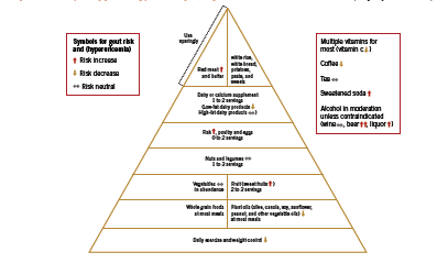
With respect to lifestyle recommendations for gout, Saag and Choi argue that the benefits of a “rigid purine restricted diet” (Saag 2006) must be weighed against the fact that a low-purine diet is often high in refined carbohydrates and saturated fat, potentially aggravating the underlying metabolic factors. Designing a diet that assists patients with weight loss, improves insulin sensitivity and lipid levels, and lowers uric acid is crucial in controlling gout symptoms as well as in managing cardiovascular risk. To this end, Choi has developed a “food pyramid” that incorporates both moderate restriction of purine intake, as well as strategies to improve insulin resistance that are characteristic of the diet used to control metabolic syndrome (2010). An adaptation of this pyramid is shown in Figure 1.

There are some notable inclusions in this dietary plan, namely, moderate amounts of purine rich legumes and vegetables, as well as fish and poultry. Choi argues that plant source purines do not increase risk of gout (2010). In a large observational study of over 47 thousand men, consumption of meat (RR 1.41, 95% CI 1.07-1.86) and seafood (RR 1.51 (1.17-1.95) but not purine rich vegetables and legumes were associated with risk of gout (Choi 2004). Increasing intake of dairy products was associated with reduced risk of gout, RR 0.56 (0.42-0.74). Choi proposes that “it would be difficult to justify a recommendation to avoid all fish intake” (2010) given its benefit on cardiovascular health, however, given the high-purine content of fish, a fish oil supplement (no protein content) seems a better strategy for patients with gout. Soft drinks and sweetened juices are discouraged, however regular consumption of coffee is allowable. Caffeine is a natural xanthine oxidase inhibitor; therefore as long as intake is consistent on a day-to-day basis, coffee may help lower uric acid levels. If intake is variable, however, coffee may in theory worsen gout, since this pattern results in unsteady changes in serum uric acid that may precipitate an attack (2010). Finally, adequate water intake is necessary to ensure uric acid excretion.

Specific Foods and Nutrients

Dairy foods An RCT recently compared the acute effect of four different kinds of milk on serum urate (Dalbeth 2010). Sixteen healthy participants received a single dose containing 80g protein of each of the following: soy milk (control); early season skim milk; late season skim milk, which contains high concentrations of orotic acid, a naturally occurring uricosuric agent; and ultrafiltrated skim milk. Soy increased serum urate approximately 10%, while the other three milks decreased urate approximately 10% (p<0.0001). Authors concluded that the urate-lowering effect of milk was due to its low purine content as well as its ability to increase urinary excretion of uric acid. In addition, certain dairy fractions including glyco-macropeptide and a milk fat extract possess anti-inflammatory properties in experimental models of acute gout (Dalbeth 2011).

Cherries A trial investigating the effect of cherry consumption was conducted in 10 healthy women (Jacob 2003). Subjects consumed two servings (280 g) of sweet cherries after an overnight fast, followed by periodic blood and urine testing. At 5h after cherry consumption, plasma urate decreased to 183 +/-15 micromol/L, compared to 214 +/-13 micro mol/L at baseline (P < 0.05). Urinary excretion of urate increased following cherries, with peak excretion of 350 +/-33 micro mol/mmol creatinine, compared to 202 +/-13 at baseline (P < 0.01).

Black tea/ Coffee In an RCT, three cups of black tea per day resulted in a significant decrease in serum uric acid such that those with baseline levels > 6 mg/dL had a 8.5% decrease (p < 0.05); while those with serum uric acid > 7 mg/dL had a decrease of 9.4% (men) and 7.1% (women) (Bahorun 2010). Coffee has been associated with decreased risk of gout, RR 0.41 (95% CI 0.19-0.88) for six or more cups per day (Choi 2007), as is expected based on its inhibition of xanthine oxidase. Since diuretic agents are known to aggravate gout, it is advisable to recommend a concomitant increase in water intake to patients consuming diuretic beverages in order to support adequate renal clearance.

Eicosapentanoic acid Although no human trials have investigated the effects of fish-derived omega-3 fatty acids on serum uric acid or risk of gout, there is good rationale for recommending EPA to gout patients on the basis of its cardioprotective and anti-inflammatory effects (Choi 2010). Studies in animal models of gout have shown that a combination of EPA and GLA reduced monosodium urate crystal-induced inflammation (Tate 1988).

Vitamin C Vitamin C is a relatively well recognized uricosuric agent, meaning that it promotes urinary excretion of uric acid. A recent meta analysis of 13 RCTs including 556 subjects and a median vitamin C dose of 500 mg/day found that there was a significant reduction in serum uric acid of -0.35 mg/dl (95%CI -0.66 to -0.03, p=0.032); in SI units this was equal to -20.8 μmoles/L (Juraschek 2011). Among trials that were placebo controlled, there were larger reductions in uric acid. Baseline serum uric acid values ranged from 2.9-7.0 mg/dl or 172.5- 416.4 μmoles/L.

Role of Food Sensitivities Although there is no reports in the literature on the association between food sensitivities and uric acid/ gout, the inflammatory state created by an immune reaction to food may aggravate gout-related inflammation and joint pain. For the case mentioned in the introduction, gluten sensitivity seems to have been an important contributing factor to the patient’s gout symptoms. These resolved dramatically on a gluten free diet combined with a selection of the other recommendations discussed here, and notably, subsequent re-exposure to gluten – while continuing other interventions, exercise, and dietary guidelines – led to a clear aggravation of the patient’s gout.

Figure 1 A healthy eating guideline pyramid for gout; impact of various foods on uric acid levels (Adapted from Choi 2010)

A Note on Drugs Because patients susceptible to gout generally have other cardiovascular risk factors such as hypertension and dyslipidemia, they are often given diruetics and/ or low-dose aspirin; however these medications may potentially precipitate gout. In a prospective study of 5789 hypertensive patients, those treated with a thiazide or loop diuretic had significantly increased risk of developing gout; HR for use of any diuretic 1.48 (95% CI 1.11-1.98), thiazide diuretic use HR 1.44 (95% CI 1.00- 2.10), and loop diuretic HR 2.31 (95% CI 1.36-3.91) (Demarco 2011). This relationship between diuretics and gout is well recognized (Saag 2006). Aspirin has been found to have a bimodal effect on excretion of uric acid: while at high doses (>3g/d) it promotes uric acid excretion (uricosuric action), at low doses (1-2g/d) it has been shown to increase retention of uric acid (Saag 2006). In another study, 75mg aspirin per day led to a 15% decrease in the rate of uric acid excretion (p=0.045) and increase in serum uric acid (p=0.009) (Caspi 2000, Saag 2006).

Conclusion Gout strongly impacts quality of life in affected patients. Because hyperuricemia is associated with the metabolic syndrome and cardiovascular risk factors, dietary strategies for gout need to be balanced in order to reduce purine intake but also reduce risk of cardiovascular disease. Weight loss is key in lowering uric acid levels. Therapeutic foods and nutritional supplements for gout include low fat dairy, black tea/ coffee, cherries, EPA, and vitamin C. The potential role of food sensitivities and medications should also be considered.

References

Bahorun T, Luximon-Ramma A, Gunness TK, Sookar D, Bhoyroo S, Jugessur R, Reebye D, Googoolye K, Crozier A, Aruoma OI. Black tea reduces uric acid and C-reactive protein levels in humans susceptible to cardiovascular diseases. Toxicology. 2010 Nov 28;278(1):68-74.

Birketvedt GS, Aaseth J, Florholmen JR, Ryttig K. Long-term effect of fibre supplement and reduced energy intake on body weight and blood lipids in overweight subjects. Acta Medica (Hradec Kralove). 2000;43(4):129-32.

Caspi D, Lubart E, Graff E, Habot B, Yaron M, Segal R. The effect of mini-dose aspirin on renal function and uric acid handling in elderly patients. Arthritis Rheum. 2000 Jan;43(1):103-8.

Cigolini M, Targher G, Tonoli M, Manara F, Muggeo M, De Sandre G. Hyperuricaemia: relationships to body fat distribution and other components of the insulin resistance syndrome in 38-year-old healthy men and women. Int J Obes Relat Metab Disord. 1995 Feb;19(2):92-6.

Choi HK. A prescription for lifestyle change in patients with hyperuricemia and gout. Curr Opin Rheumatol. 2010 Mar;22(2):165-72.

Choi HK, Willett W, Curhan G. Coffee consumption and risk of incident gout in men: a prospective study. Arthritis Rheum. 2007 Jun;56(6):2049-55.

Choi HK, Atkinson K, Karlson EW, Curhan G. Obesity, weight change, hypertension, diuretic use, and risk of gout in men: the health professionals follow-up study. Arch Intern Med. 2005 Apr 11;165(7):742-8.

Choi HK, Atkinson K, Karlson EW, Willett W, Curhan G. Purine-rich foods, dairy and protein intake, and the risk of gout in men. N Engl J Med. 2004 Mar 11;350(11):1093-103.

Dalbeth N, Wong S, Gamble GD, Horne A, Mason B, Pool B, Fairbanks L, McQueen FM, Cornish J, Reid IR, Palmano K. Acute effect of milk on serum urate concentrations: a randomised controlled crossover trial. Ann Rheum Dis. 2010 Sep;69(9):1677-82.

Dalbeth N, Palmano K. Effects of dairy intake on hyperuricemia and gout. Curr Rheumatol Rep. 2011 Apr;13(2):132-7.

Demarco MA, Maynard JW, Baer AN, Gelber AC, Young JH, Alonso A, Coresh J. Diuretic use, increased serum urate and the risk of incident gout in a populationbased study of hypertensive adults: The atherosclerosis risk in the communities cohort. Arthritis Rheum. 2011 Sep 27. doi: 10.1002/art.33315.

Emmerson BT. The management of gout. N Engl J Med. 1996 Feb 15;334(7):445-51.

Gibson T. Hyperuricemia, gout and the kidney. Curr Opin Rheumatol. 2011 Dec 12. [Epub ahead of print]

Inokuchi T, Tsutsumi Z, Takahashi S, Ka T, Moriwaki Y, Yamamoto T. Increased frequency of metabolic syndrome and its individual metabolic abnormalities in Japanese patients with primary gout. J Clin Rheumatol. 2010 Apr;16(3):109-12.

Jacob RA, Spinozzi GM, Simon VA, Kelley DS, Prior RL, Hess-Pierce B, Kader AA. Consumption of cherries lowers plasma urate in healthy women. J Nutr. 2003 Jun;133(6):1826-9.

Juraschek SP, Miller ER 3rd, Gelber AC. Effect of oral vitamin C supplementation on serum uric acid: a meta-analysis of randomized controlled trials. Arthritis Care Res (Hoboken). 2011 Sep;63(9):1295-306.

Johnson WD, Brashear MM, Gupta AK, Rood JC, Ryan DH. Incremental weight loss improves cardiometabolic risk in extremely obese adults. Am J Med. 2011 Oct;124(10):931-8.

Krzystek-Korpacka M, Patryn E, Kustrzeba-Wojcicka I, Chrzanowska J, Gamian A, Noczynska A. The effect of a one-year weight reduction program on serum uric acid in overweight/obese children and adolescents. Clin Chem Lab Med. 2011 May;49(5):915-21.

Madero M, Arriaga JC, Jalal D, Rivard C, McFann K, Pérez-Méndez O, Vázquez A, Ruiz A, Lanaspa MA, Jimenez CR, Johnson RJ, Lozada LG. The effect of two energyrestricted diets, a low-fructose diet versus a moderate natural fructose diet, on weight loss and metabolic syndrome parameters: a randomized controlled trial. Metabolism. 2011 Nov;60(11):1551-9.

Meshkani R, Zargari M, Larijani B. The relationship between uric acid and metabolic syndrome in normal glucose tolerance and normal fasting glucose subjects. Acta Diabetol. 2011 Mar;48(1):79-88.

Pascual E, Sivera F. Therapeutic advances in gout. Curr Opin Rheumatol. 2007 Mar;19(2):122-7.

Rathmann W, Funkhouser E, Dyer AR, Roseman JM. Relations of hyperuricemia with the various components of the insulin resistance syndrome in young black and white adults: the CARDIA study. Coronary Artery Risk Development in Young Adults. Ann Epidemiol. 1998 May;8(4):250-61.

Rx List Online. Zyloprim (Allopurinol). Updated 2011. http://www.rxlist.com/ zyloprim-drug.htm Accessed December 22 2011.

Saag KG, Choi H. Epidemiology, risk factors, and lifestyle modifications for gout. Arthritis Res Ther. 2006;8 Suppl 1:S2.

Smith HS, Bracken D, Smith JM. Gout: current insights and future perspectives. J Pain. 2011 Nov;12(11):1113-29.

Tate GA, Mandell BF, Karmali RA, Laposata M, Baker DG, Schumacher HR Jr, Zurier RB. Suppression of monosodium urate crystal-induced acute inflammation by diets enriched with gamma-linolenic acid and eicosapentaenoic acid. Arthritis Rheum. 1988 Dec;31(12):1543-51.

Yamada T, Fukatsu M, Suzuki S, Wada T, Joh T. Elevated serum uric acid predicts impaired fasting glucose and type 2 diabetes only among Japanese women undergoing health checkups. Diabetes Metab. 2011 Jun;37(3):252-8.

Drug-free blood pressure control advice found in an online self-help kit

0

Stress management psychologist, Yehudit Feuer, Ph.D., developed the Brelax[TM] method for the treatment of hypertension. It includes several home-based programs for enhanced mind-body wellness, based on elements of classical yoga and stress management techniques. All of the Brelax programs consist mainly of moderate exercises coordinated with deep breathing and systematic relaxation, which makes them aerobic by definition and applicable across a wide age range. One of these programs that focused specifically on cardiovascular disorders was hospital- tested for effectiveness with Brelax- trained patients in two controlled studies. In hypertensive patients, the treatment group experienced a significant reduction in average blood pressure values, and in patients with coronary heart disease, cardiac performance and fitness improved significantly, independent of other life-style changes. The training is now available online (http://www.brelaxcenter. com) as a self-help kit. Two richly illustrated user manuals explain the theory behind the method and two constantly available guides on compact disks are available to facilitate regular practice.

The Government of Canada requests research proposals for clinical trial on CCSVI and MS

0

The Honourable Leona Aglukkaq, Minister of Health, and Dr. Alain Beaudet, President of the Canadian Institutes of Health Research (CIHR) have announced that CIHR is ready to accept research proposals for the Phase I/ II clinical trial on Chronic Cerebrospinal Venous Insufficiency (CCSVI). The request for research proposals was available on CIHR’s website on November 30, 2011 and CIHR plans to announce the successful research team in March 2012. “We are excited to collaborate with CIHR in bringing the multiple sclerosis (MS) community closer to definitive answers on CCSVI and MS,” said Yves Savoie, President and CEO of the MS Society. “People with MS deserve clarity about the hope that CCSVI offers as a potential treatment for MS. It is only through rigorous research that we can get theseanswers.”

Tabatha Parker was chosen as an Utne Reader visionary in 2011

0

Each year, Utne Reader puts forward its selection of world visionaries and has chosen Tabatha Parker, ND, founder of Natural Doctors International (NDI). “The global health care system is in crisis,” says Parker. She notes that it relies on the exportation of a Western model to developing countries that can’t afford it. Parker sees NDI as a bridge between exported conventional medicine and centuries-old indigenous healing techniques, such as the use of herbal medicine, which in some places in the world is the dominant type of health care. “That has to be a part of the system if you’re going to actually reach people,” Parker says. Naturopathic doctors “are trained in a way that no one else in the world is trained: to be [that] bridge.” NDI has a location in Nicaragua and it has worked with the World Health Organization’s traditional medicine arm to broaden its understanding of natural medicine. Parker sees NDI becoming a sort of Doctors Without Borders for naturopaths and she hopes to establish permanent sites worldwide that all serve as a link between old and new.

Become a member of the Seniors Health Research Transfer Network’s Nutrition Community of Practice

0

The Seniors Health Research Transfer Network (SHRTN) has been operating a Nutrition Community of Practice (CoP) for the past year that has the following goals: 1) link current and future practitioners involved in the nutrition care of older adults; 2) promote evidence-based nutrition care for older adults by supporting knowledge exchange among experts, researchers, professional organizations, practitioners, and caregivers; 3) disseminate best practices in nutrition care to frontline caregivers; 4) advocate for innovation and best practices in the nutrition care of older adults; and 5) stimulate research on food and nutrition issues for older adults. The CoP hosted a webinar in Spring 2011 titled ‘Making the Most of Mealtimes’ and every month, reading lists on a number of topics related to nutrition and older adults are disseminated to CoP members. To become a member and access the CoP network and resources, visit the SHRTN website at http://beta.shrtn.on.ca.

A new web-based service will transform the way health care providers and patients collaborate and exchange information

0

At the Ontario Hospital Association’s HealthAchieve 2011 Conference, McKesson Canada announced the launch of RelayHealth, a new web-based service that provides complete and secure information flow between patients and all of their health care providers from coast to coast. RelayHealth is intended to be made available at no cost to patients and will accelerate information exchange, facilitate collaboration between health care providers, advance patient quality of care, and reduce overall health care costs. It will provide patients with the tools they need to better manage their own health by allowing physicians, health care providers, and patients to share lab and diagnostic test results, make referral requests, book appointments, make prescription and refill requests, and reduce duplicate testing and medical errors. Dale Weil of McKesson Canada says, “The advantage of using this service is that it achieves savings in the shorter term for the health care system and it will help increase patient satisfaction. Also, because RelayHealth is delivered as a service, it accelerates implementation at a lower cost than other major health information technologies and it requires less maintenance. It is a convenient service that provides the same view, same patient information across the entire health network.”

Coalition against morbid obesity launches “Combat Morbid Obesity” campaign

0

The Coalition against Morbid Obesity has launched a social media campaign to combat morbid obesity in an attempt to break the isolation of people who are morbidly obese, raise their awareness of the dangerous effects of their disease on their health and quality of life, and inform them about the existence of an effective treatment. In two video testimonies that can be found on YouTube and the Coalition’s Website (www.lepoidsquitue. com) and Facebook page, women speak about their experiences with this condition. In the first video, a woman talks about becoming aware of the fact that she could not fight this disease alone and about the help she sought out. In the second video, another woman talks about her decision to undergo bariatric surgery in 2004, which was a choice she made to improve her quality of life. In the campaign’s second stage, people with morbid obesity will be invited to create their own video message. The Coalition hopes that this campaign will go viral to encourage people with morbid obesity to learn more about the disease, about the dangers it presents to their health and quality of life, and about bariatric surgery.

Canada’ s pioneer of integrative cancer care passes on

0

Dr. Roger Rogers, BA, BSW, MD, OBC, died peacefully on November 22, 2011 at the age of 83 years. As a co-founder of InspireHealth, Canada’s first integrative cancer care centre, Dr. Rogers is recognized as a pioneer and leader in integrative cancer care. “It is with sadness and the celebration of a life well lived that I share, with the extended InspireHealth family, that Dr. Rogers has peacefully passed away. Loved by his patients as much as he loved them, Roger was an inspiration to several generations of physicians,” commented Dr. Hal Gunn, co-founder and CEO of InspireHealth. Dr. Rogers was the grateful recipient of many awards and acknowledgements, culminating in the presentation of the Order of British Columbia in 2001. The Hecht Foundation created the Dr. Roger Rogers Prize in 2007, which awards a $250 000 prize biennially to a leader in complementary and alternative medicine. Dr Rogers leaves the legacy of an extraordinary career that will continue to shape and influence the future of cancer care not only in Canada but also worldwide.