




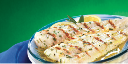
This study examined the associations between dietary polyunsaturated fatty acid (PUFA) and fish intakes and hip bone mineral density (BMD) in adults (n = 623; mean age of 75 years) in the Framingham Osteoporosis Study at baseline and four years later. High intakes (≥3 servings/wk) of fish relative to lower intakes were associated with maintenance of femoral neck BMD (FN-BMD) in men (dark fish + tuna, dark fish, and tuna) and women (dark fish) (P < 0.05). Significant interactions between arachidonic acid (AA) and eicosapentaenoic acid (EPA)+ docosahexaenoic acid (DHA) intakes were observed cross-sectionally in women and longitudinally in men. In women with EPA+DHA intakes at or above the median, those with the highest AA intakes had a higher mean baseline FN-BMD than those with the lowest intakes (P = 0.03, P for trend = 0.02). In men with the lowest EPA+DHA intakes, those with the highest intakes of AA lost more FN-BMD than men with the lowest intakes of AA (P = 0.04). The authors concluded that fish consumption may protect against bone loss and the protective effects of a high AA intake may be dependent on EPA+DHA intake. Am J Clin Nutr. 2011 May;93(5):1142-51. PMID: 21367955
Mulberry leaf tablets improve cholesterol in patients with mild dyslipidemia
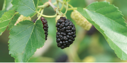
A within-subjects research design was conducted at an out-patient clinic in Thailand to evaluate the hypolipidemic effects of mulberry leaf in non-diabetic patients with mild dyslipidemia.. Twenty-three patients who met the National Cholesterol Education Program Adult Treatment Panel III criteria guideline for dyslipidemia and failed a four-week diet therapy were enrolled. Subjects were assigned to receive three tablets of 280 mg mulberry leaf per tablet three times daily before meals for 12 weeks and blood analyses were performed every four weeks. At four and eight weeks of mulberry leaf tablet therapy, triglycerides (TG) had significantly decreased by 10.2% (p < 0.05) and 12.5% (p < 0.05), respectively, from baseline. At the end of the study, total cholesterol, TG, and low-density lipoprotein cholesterol had decreased by 4.9% (p < 0.05), 14.1% (p < 0.05), and 5.6% (p < 0.05), respectively, from baseline, whereas high-density lipoprotein (HDL) cholesterol had increased by 19.7% (p < 0.05). Although some patients experienced such side effects as mild diarrhea (26%), dizziness (8.7%), or constipation and bloating (4.3%), mulberry leaf tablet therapy is considered safe and effective in decreasing cholesterol and increasing HDL levels in patients with mild dyslipidemia. Phytother Res. 2011 Mar;25(3):365-9. PMID: 20687135
The impact of comorbid cannabis use disorders on the clinical presentation of social anxiety disorder

Cannabis use disorders (CUDs) are becoming increasingly problematic within the population of individuals with social anxiety disorder (SAD), yet the nature of this comorbidity remains largely unexamined. The aim of the current study from the Rhode Island Methods to Improve Diagnostic Assessment and Services (MIDAS) project was to examine differences between SAD outpatients with versus without comorbid CUDs. Patients with SAD and comorbid CUDs (n = 173) were compared to those with SAD without CUDs (n = 700) on demographic and clinical characteristics. Compared to patients without the comorbidity, patients with comorbid SAD and CUDs were more likely to have a lifetime diagnosis of post-traumatic stress disorder, specific phobias, lifetime substance use disorders (including alcohol), and report better physical health and fewer limitations related to their physical health. These analyses remained significant after controlling for gender, the presence of other substance use disorders, mood disorders, and other anxiety disorders. Findings of this study suggest that there may be a unique relationship between SAD and CUDs that can potentially impact the clinical presentation of individuals with SAD. Future research is needed to examine the impact of this comorbidity in other patient populations. J Psychiatr Res. 2011 Oct 31. PMID: 22047609
Efficacy of a web-based food and exercise diary in a commercial weight loss program

A retrospective analysis was conducted to assess weight change among 3621 subscribers (2979 women; 642 men) to a commercial internet-based weight loss program. Program engagement (indexed with frequency of using online diet and exercise diaries and with use of the social support forums) was associated with weight loss in both men and women after controlling for initial BMI and duration of participation. These engagement variables accounted for 13% of variance in percentage weight loss in women (p<0.001) and 19% in men (p<0.001). Exercise diary use predicted weight loss among men but not women and the use of online forums predicted weight loss among women but not men. Among participants who were overweight or obese, those in the highest tertile of engagement with food diaries (vs the lowest) were more likely to achieve clinically significant (>5%) weight loss (p<0.001 for both men and women). Being in the highest tertile of engagement with exercise diaries was associated with clinically significant weight loss in men (p<0.001) and, less strongly, in women (p<0.05). These results suggest that the use of self-monitoring tools and participation in online support are predictive of weight loss. Int J Behav Nutr Phys Act. 2011 Aug 2;8:83. PMID: 21810222
Reduction of common cold symptoms by encapsulated juice powder concentrate of fruits and vegetables

Nutraceuticals have shown benefit in the prevention of the common cold but previous investigations have been inconsistent. This study was designed to determine the preventive effect of a dietary supplement from fruits and vegetables on common cold symptoms. In a double-blind, placebo-controlled trial, 529 healthcare professionals (mean age 39.9 years) from a university hospital in Germany were randomized to receive four capsules of a dietary supplement (Juice Plus+®) or a matching placebo daily for eight months. The number of days with moderate or severe common cold symptoms within six months was assessed by diary self-reports. The mean number of days with moderate or severe common cold symptoms was 7.6 (95 % CI 6.5 – 8.8) in the Juice Plus+® group and 9.5 (95% CI 8.4 – 10.6) in the placebo group (p = 0.023). The mean number of total days with any common cold symptoms was similar between the Juice Plus+® and placebo groups. The authors concluded that the intake of a dietary supplement from fruits and vegetables was associated with a 20% reduction of moderate or severe common cold symptom days in healthcare professionals particularly exposed to patient contact. Br J Nutr. 2011 Jan;105(1):118-22. PMID: 20727236
Early-life bisphenol A exposure negatively affects behavior and emotional regulation in children.

A prospective birth cohort of 244 mothers and their three-year-old children from Ohio was conducted to estimate the impact of gestational and childhood bisphenol A (BPA) exposures on behavior and executive function at three years of age. BPA concentrations were measured in >97% of maternal (16 and 26 weeks of gestation and birth; median 2.0 μg/L) and child (1, 2, and 3 years of age; median 4.1 μg/L) urine samples. With adjustment for confounders, each 10-fold increase in gestational BPA concentrations was associated with more anxious and depressed behavior on the Behavior Assessment System for Children 2 (BASC-2) and poorer emotional control and inhibition on the Behavior Rating Inventory of Executive Function-Preschool (BRIEF-P). The magnitude of the gestational BPA associations differed according to child gender; BASC-2 and BRIEF-P scores increased 9 to 12 points among girls but changes were null or negative among boys. Associations between childhood BPA exposure and neurobehavior were largely null and not modified by child gender. The authors concluded that gestational BPA exposure affected behavioral and emotional regulation domains, especially among girls and that clinicians may want to advise concerned patients to reduce their exposure to certain consumer products. Pediatrics. 2011 Nov;128(5):873-82. PMID: 22025598
Whey protein may decrease inflammation and increase antioxidant defenses in elderly patients with ischemic stroke

This randomized study compared the effects of an early enteral formula containing whey protein to a standard enteral formula containing casein on the levels of glutathione and inflammatory markers in aged patients with acute ischemic stroke. Thirty-one elderly patients (12 males and 19 females; median age 74 years) with ischemic stroke were randomized to receive early nasogastric feeding (35 kcal/kg/day and 1.2 g of protein/kg/day) with either a formula containing hydrolyzed casein or another isocaloric and isonitrogenous formula containing hydrolyzed whey protein for five days. Mortality was found to be similar between groups and was associated with higher serum IL-6 (p = 0.04) and C-reactive protein (p = 0.02) levels. Serum IL-6 decreased (p = 0.02) and glutathione increased (p = 0.03) only in the whey protein group. In addition, serum IL-6 was lower (p = 0.03) and glutathione was higher (p = 0.03) in the whey protein group compared to the casein group. The authors concluded that an early-administered whey protein formula seems to be more effective than a casein formula at decreasing inflammation and increasing antioxidant defenses in elderly patients with ischemic stroke. Nutrition. 2011 Apr;27(4):440-4. PMID: 21167685
Acupuncture is effective for the treatment of severe acute pain in Herpes Zoster

The current study was conducted to assess the potential efficacy of acupuncture in controlling intense or very intense pain in patients with Herpes Zoster in comparison with standard pharmacological treatment. One hundred and two patients were randomized to receive either acupuncture (n=52) or standard pharmacological treatment (n=50) for four weeks. Response rates, mean changes in pain intensity, differences in total pain burden with an area-under-the-curve (AUC) method over a one-year follow-up, and differences in the incidence of Post-Herpetic Neuralgia (PHN) were evaluated. Both interventions were largely effective. No significant differences were observed in response rates (81.6% vs 89.2%; p = 0.8), mean reduction of visual analogue scale (4.1+/-2.3 vs 4.9+/-1.9; p = 0.12) and McGill Pain Questionnaire scores (1.3+/-0.9 vs 1.3+/-0.9; p = 0.9), incidence of PHN after three months (48.4% vs 46.8%; p = 0.5), and mean AUC during follow-up (199+/-136 vs 173+/-141; p = 0.4). No serious treatmentrelated adverse events were observed in both groups. The authors concluded that this controlled and randomized trial provides the first evidence of a potential role of acupuncture for the treatment of acute herpetic pain. BMC Complement Altern Med. 2011 Jun 5;11(1):46. PMID: 21639941
Pregnancy Morbidity

Pregnancy Morbidity
Update on Antiphospholipid Syndrome (APS)
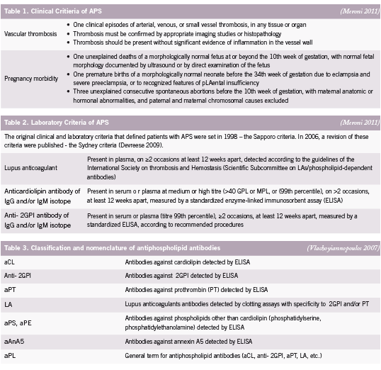
e antiphospholipid syndrome (APS) is an autoimmune disease de ned by two major elements: the occurrence of clinical features, categorised as recurrent vascular thrombosis and/or pregnancy morbidity, and the presence of antiphospholipid antibodies (aPL) in plasma (Devreese 2009). Antiphospholipid syndrome is sometimes referred to as Hughes syndrome, after the rheumatologist Dr. Graham R.V. Hughes. Clinical criteria for APS includes pregnancy loss – either as miscarriage, intrauterine fetal death or stillbirth – and/or obstetric complications, such as intrauterine growth restriction (IUGR), uteroplacental insu ciency, pre-eclampsia and pre-term birth (before 30 weeks) (Carp 2008, Yasuda 1995) (See Table 1). Signi cant associations between recurrent miscarriages and pregnancy complications have been reported with each autoantibody, detectable by di erent assays, in the aPL family including lupus anticoagulant (LA), anti-cardiolipin (aCL) and anti-beta2 glycoprotein I (β2GPI) (Tincani 2003) (See Table 2). Assays of the various aPL are not only diagnostic of APS, but are believed to have a pathogenic role, mediating several clinical manifestations of the syndrome (Meroni 2010).
Antibody Measurements in APS
Antiphospholipid syndrome was rst identi ed more than 20 years ago when the lupus anticoagulant (LA), known to prolong prothrombin time (PT), was found in patients with a history of thrombosis and recurrent second trimester pregnancy loss (Tincani 2003). It is currently considered more appropriate to assess a combination of antibodies rather than one antibody for adequate laboratory detection of APS as no antibody is pathognomic for pregnancy loss or reproductive complications (Carp 2008) (See Table 3).
LA recognition in particular has several disadvantages. Handling and preparation of plasma samples is complicated and labourintensive (Devreese 2009).
e diagnostic value of the aCL antibody test is currently under debate, partly because of methodological problems (Devreese 2009); however, aCL have been shown to help predict cases of Systemic lupus erythematosus (SLE) shifting to secondary APS (Harel 2006).
Antibodies against β2GPI seem to be particularly important, especially when aCL and LA are negative and APS is strongly suspected (Bas de Laat 2004, de Groot 2005). In 3% to 10% of APS patients, anti-β2GPI antibodies might be the only positive test (Ebeling 2003, Lee 2003, Nash 2004). e anti-β2GPI antibody ELISAs (Enzyme-linked immunosorbent assay) detect all antibodies reactive with the protein, making it less speci c, and probably not suitable as a general diagnostic test (de Laat 2008).
Combining the pro le for all three of the tests may enable correct interpretation of results and clinical management (Pengo 2007). A full positive pro le (ie. presence of LA, aCL and β2GPI antibodies) is associated with thrombosis and obstetric complications and appears to re ect the presence of large amounts of antibodies speci cally against β2GPI. LA shows the strongest correlation with thrombosis and pregnancy morbidity (Galli 2003). Patients positive for aCL and β2GPI antibodies, with no history of thromboembolic complications, presented only with obstetric complications (Pengo 2005, Ru atti 2006).
Pathogenic Mechanisms
The thrombophillic e ect of aPL in intraplacental thrombosis, impairing maternal-fetal blood exchange, was originally thought to be the main pathogenic mechanism of fetal loss (Meroni 2010). Histopathological ndings failed to nd thrombosis as the cause of all reproductive failure in samples of miscarried fetuses and placentas from women with APS however (Park 2006). Though thrombosis is indeed a part of the pathophysiology, it is well recognized that different mechanisms are responsible for the vascular and the obstetrical manifestations of APS (Meroni 2010).
Thrombosis
In vivo models of thrombosis induced in mice and hamsters have confirmed that aPL are able to increase thrombus formation in venous and arterial trees (Fischetti 2005, Jankowski 2003,Ramesh 2011). Infusion with and without β2GPI revealed altered expression of endothelial adhesion molecules, and an upregulated expression of nitric oxide and tissue factor (TF) have been reported in arterial endothelia, supporting a key role for aPL in causing vascular abnormalities (Ramesh 2011, Romay- Penabad 2011, Vega-Ostertag 2007). It is the β2GPI, of all the aPL subpopulations, that appears to be most responsible for the thrombotic manifestations of APS (Meroni 2010).
Impaired Trophoblast Invasiveness
Trophoblast invasion is a dynamic process that is tightly controlled by a complex series of interactions between trophoblast and decidual tissues; defective placentation may occur as a result of default in any of them (Meroni 2010, Pierangeli 2008). The differentiation of the trophoblast into an invasive phenotype is related to the expression of cell surface adhesion and signalling molecules (Aplin 2000, Castellucci 2000). Placental invasion might then be affected by abnormal integrin and cadherin expression caused by aPL activity (Di Simone 2002). The failure to express the correct adhesion molecule phenotype by the trophoblast is the disruption that is thought to prevent proper decidual invasion (Meroni 2004). These mechanisms have been suggested to play a role in early fetal loss, while thrombotic events would be responsible for miscarriages late in pregnancy (Meroni 2004).
Uncontrolled Inflammatory Balance
Evidence for the delicate balance between proinflammatory and anti-inflammatory mediators during gestation on the outcome of pregnancy exist (Chaouat 2007). Proinflammatory mediators (such as complement, tumor necrosis factor [TNF], and CC chemokines) have been shown to have a role in animal models of aPL-induced fetal loss (Meroni 2010).
Endogenous Protective Mediator
Annexin V or placental anti-coagulant protein I (PAP-I) is a protein that is expressed on the surface of villous syncitiotrophoblasts. It displays a strong anticoagulant activity in APS by binding to the negatively charged phospholipids, thereby producing a protective shield against aPLs that would otherwise bind to these same phospholipids (Tincani 2003). In APS patients, a decreased quantity of Annexin V has been reported at the placental level (Krikun 1994).
Conventional Treatment of APS
In spite of the general consensus on the management of pregnant aPL-positive women, few well-designed clinical trials have been reported and there is also insufficient data about the positive predictive value (PPV) of treatment (Carp 2008). To date, concomitant administration of heparin and aspirin are used in the treatment of APS. This followed a 1992 controlled trial using 20,000 U/day heparin and 81 mg aspirin that revealed improved pregnancy outcome (Cowchock 1992). The rationale for heparin use was originally founded on the pathogenic role of thrombotic phenomena in aPL-associated pregnancy loss (Hughes 1983), but it was the anticomplement, rather than the anticoagulant, effect of heparin that was later credited in experimental models with mice for the positive pregnancy outcomes (Girardi 2004).
Aspirin has been shown to stimulate Interleukin-3 (IL-3) production (Fishman 1995). IL-3 is a known growth factor for the trophoblast (Wegmann 1989) and IL-3 levels are reduced in women diagnosed with APS during pregnancy (Fishman 1996). In vivo studies in experimental models revealed compelling results such as the elimination of aPL-related obstetrical complications through the addition of exogenous IL-3 (Fishman 1993) that upheld the continued use of aspirin in APS. Some in vitro studies however were unable to provide evidence that IL-3 prevented aPL effect on the trophoblast (Chamley 2001), while others could (Di Simone 2000).
Low corticosteroid doses (<20 mg/day) are occasionally used, particularly in women unresponsive to the standard low-dose aspirin and heparin therapy (Meroni 2011), but there is no sound evidence to support the routine use of corticosteroids in APS (Cowchock 1992, Ruiz-Irastorza 2010). Intravenous immunoglobulin use as a prophylactic treatment of fetal loss in APS patients has been discussed (Tincani 2003). This treatment, added to conventional heparin/aspirin treatment, was unable to improve reproductive prognosis in APS patients (Branch 2000). It is thought that such treatment should be reserved for cases in which conventional therapy has failed or to treat acute problems of pregnancy, especially acute problems of pregnant APS patients (Tincani 2003).
Populations at Greatest Risk for APS
The link between the presence of anti-thyroid antibodies and either isolated or recurrent pregnancy loss has been reproducibly demonstrated (Bussen 1995, Glinoer 1991, Singh 1995, Stagnaro-Green 1990). Presence of anti-thyroid antibodies are significantly correlated with an increased rate of miscarriage in women in their first trimester of pregnancy (Stagnaro-Green 1990) and an increased rate of spontaneous pregnancy loss was reported in women with detectable thyroid autoantibodies compared with controls (Glinoer 1991). Approximately one-third of women diagnosed with APS had anti-thyroid antibodies in a study by De Carolis and colleagues. Women with anti-thyroid antibodies (associated or not with aPL) had significantly lower percentages of pregnancies, and the overall success rate of pregnancy in patients positive for thyroid autoimmunity (alone or association with aPL) was 59% (De Carolis 2004). The presence of anti-thyroid antibodies have an overall PPV of 40% for pregnancy loss (Carp 2008).
That women with systemic lupus erythematosus (SLE) are at greater risk of experiencing pregnancy loss is nothing new. SLE has been known for decades to predispose to spontaneous abortions (Gleicher 1999). It is now known, however, that the risk for pregnancy loss precedes the clinical occurrence and/or diagnosis of SLE as well (Hardy 1999). This bidirectional phenomena is termed reproductive autoimmune failure syndrome (RAFS) and is thought to extend to autoimmune conditions beyond SLE (Gleicher 1999). Many authors suggested that, in addition to other causes, infertile patients might suffer from subclinical autoimmune disease and, in turn, those subclinical autoimmune patients could be at risk of implantation failure or of early post-implantation loss (Geva 1997).
References
Aplin JD, Haigh T, LAey H, Chen CP and Jones CJ. Tissue interactions in the control of trophoblast invasion. J Reprod Fertil Suppl. 2000; 55 57-64.
Bas de Laat H, Derksen RH and de Groot PG. beta2-glycoprotein I, the playmaker of the antiphospholipid syndrome. Clin Immunol. 2004; 112 (2): 161-168.
Branch DW, Peaceman AM, Druzin M, Silver RK, El-Sayed Y, Silver RM, Esplin MS, Spinnato J and Harger J. A multicenter, pLAebo-controlled pilot study of intravenous immune globulin treatment of antiphospholipid syndrome during pregnancy. The Pregnancy Loss Study Group. Am J Obstet Gynecol. 2000; 182 (1 Pt 1): 122-127.
Bussen S and Steck T. Thyroid autoantibodies in euthyroid non-pregnant women with recurrent spontaneous abortions. Hum Reprod. 1995; 10 (11): 2938-2940.
Carp HJ, Meroni PL and Shoenfeld Y. Autoantibodies as predictors of pregnancy complications. Rheumatology. 2008; 47 Suppl 3 iii6-8.
Castellucci M, De Matteis R, Meisser A, Cancello R, Monsurro V, Islami D, Sarzani R, Marzioni D, Cinti S and Bischof P. Leptin modulates extracellular matrix molecules and metalloproteinases: possible implications for trophoblast invasion. Mol Hum Reprod. 2000; 6 (10): 951-958.
Chamley LW, Konarkowska B, Duncalf AM, Mitchell MD and Johnson PM. Is interleukin-3 important in antiphospholipid antibody-mediated pregnancy failure? Fertil Steril. 2001; 76 (4): 700-706.
Chaouat G. The Th1/Th2 paradigm: still important in pregnancy? Semin Immunopathol. 2007; 29 (2): 95-113.
Cowchock FS, Reece EA, Balaban D, Branch DW and Plouffe L. Repeated fetal losses associated with antiphospholipid antibodies: a collaborative randomized trial comparing prednisone with low-dose heparin treatment. Am J Obstet Gynecol. 1992; 166 (5): 1318- 1323.
De Carolis C, Greco E, Guarino MD, Perricone C, Dal Lago A, Giacomelli R, Fontana L and Perricone R. Anti-thyroid antibodies and antiphospholipid syndrome: evidence of reduced fecundity and of poor pregnancy outcome in recurrent spontaneous aborters. Am J Reprod Immunol. 2004; 52 (4): 263-266.
de Groot PG and Derksen RH. Pathophysiology of the antiphospholipid syndrome. J Thromb Haemost. 2005; 3 (8): 1854-1860.
de Laat B, Mertens K and de Groot PG. Mechanisms of disease: antiphospholipid antibodiesfrom clinical association to pathologic mechanism. Nat Clin Pract Rheumatol. 2008; 4 (4): 192-199.
Devreese K and Hoylaerts MF. Laboratory diagnosis of the antiphospholipid syndrome: a plethora of obstacles to overcome. Eur J Haematol. 2009; 83 (1): 1-16.
Di Simone N, Caliandro D, Castellani R, Ferrazzani S and Caruso A. Interleukin-3 and human trophoblast: in vitro explanations for the effect of interleukin in patients with antiphospholipid antibody syndrome. Fertil Steril. 2000; 73 (6): 1194-1200.
Di Simone N, Castellani R, Caliandro D and Caruso A. Antiphospholid antibodies regulate the expression of trophoblast cell adhesion molecules. Fertil Steril. 2002; 77 (4): 805-811.
Ebeling F, Pettersson T, Muukkonen L, Vahtera E and Rasi V. Beta-2-glycoprotein I antibodies in patients with thrombosis. Scand J Clin Lab Invest. 2003; 63 (2): 111-118.
Fischetti F, Durigutto P, Pellis V, Debeus A, Macor P, Bulla R, Bossi F, Ziller F, Sblattero D, Meroni P and Tedesco F. Thrombus formation induced by antibodies to beta2-glycoprotein I is complement dependent and requires a priming factor. Blood. 2005; 106 (7): 2340-2346.
Fishman P, Falah-Vaknin E, Sredni B, Meroni PL, Rudniki C and Shoenfeld Y. Aspirin modulates interleukin-3 production: additional explanation for the preventive effects of aspirin in antiphospholipid antibody syndrome. J Rheumatol. 1995; 22 (6): 1086-1090.
Fishman P, FaLAh-Vaknin E, Sredni B, Meroni PL, Tincani A, Dicker D and Shoenfeld Y. Aspirin-interleukin-3 interrelationships in patients with anti-phospholipid syndrome. Am J Reprod Immunol. 1996; 35 (2): 80-84.
Fishman P, FaLAh-Vaknine E, Zigelman R, Bakimer R, Sredni B, Djaldetti M and Shoenfeld Y. Prevention of fetal loss in experimental antiphospholipid syndrome by in vivo administration of recombinant interleukin-3. J Clin Invest. 1993; 91 (4): 1834-1837.
Galli M, Luciani D, Bertolini G and Barbui T. Lupus anticoagulants are stronger risk factors for thrombosis than anticardiolipin antibodies in the antiphospholipid syndrome: a systematic review of the literature. Blood. 2003; 101 (5): 1827-1832.
Geva E, Amit A, Lerner-Geva L and Lessing JB. Autoimmunity and reproduction. Fertil Steril. 1997; 67 (4): 599-611.
Girardi G, Redecha P and Salmon JE. Heparin prevents antiphospholipid antibody-induced fetal loss by inhibiting complement activation. Nat Med. 2004; 10 (11): 1222-1226.
Gleicher N. Reproductive failure prior to the onset of clinical autoimmune disease. Rheumatology. 1999; 38 (6): 485-487.
Glinoer D, Soto MF, Bourdoux P, Lejeune B, Delange F, Lemone M, Kinthaert J, Robijn C, Grun JP and de Nayer P. Pregnancy in patients with mild thyroid abnormalities: maternal and neonatal repercussions. J Clin Endocrinol Metab. 1991; 73 (2): 421-427.
Hardy CJ, Palmer BP, Morton SJ, Muir KR and Powell RJ. Pregnancy outcome and family size in systemic lupus erythematosus: a case-control study. Rheumatology (Oxford). 1999; 38 (6): 559-563.
Harel M and Shoenfeld Y. Predicting and preventing autoimmunity, myth or reality? Ann N Y Acad Sci. 2006; 1069 322-345.
Jankowski M, Vreys I, Wittevrongel C, Boon D, Vermylen J, Hoylaerts MF and Arnout J. Thrombogenicity of beta 2-glycoprotein I-dependent antiphospholipid antibodies in a photochemically induced thrombosis model in the hamster. Blood. 2003; 101 (1): 157-162.
Krikun G, Lockwood CJ, Wu XX, Zhou XD, Guller S, Calandri C, Guha A, Nemerson Y and Rand JH. The expression of the pLAental anticoagulant protein, annexin V, by villous trophoblasts: immunolocalization and in vitro regulation. PLAenta. 1994; 15 (6): 601-612.
Lee EY, Lee CK, Lee TH, Chung SM, Kim SH, Cho YS, Yoo B and Moon HB. Does the antibeta2- glycoprotein I antibody provide additional information in patients with thrombosis? Thromb Res. 2003; 111 (1-2): 29-32.
Meroni PL, Borghi MO, Raschi E and Tedesco F. Pathogenesis of antiphospholipid syndrome: understanding the antibodies. Nat Rev Rheumatol. 2011; 7 (6): 330-339.
Meroni PL, di Simone N, Testoni C, D’Asta M, Acaia B and Caruso A. Antiphospholipid antibodies as cause of pregnancy loss. Lupus. 2004; 13 (9): 649-652.
Meroni PL, Tedesco F, Locati M, Vecchi A, Di Simone N, Acaia B, Pierangeli SS and Borghi MO. Anti-phospholipid antibody mediated fetal loss: still an open question from a pathogenic point of view. Lupus. 2010; 19 (4): 453-456.
Nash MJ, Camilleri RS, Kunka S, Mackie IJ, Machin SJ and Cohen H. The anticardiolipin assay is required for sensitive screening for antiphospholipid antibodies. J Thromb Haemost. 2004; 2 (7): 1077-1081.
Park, A.L. in Hughes’ Syndrome (ed. Khamashta, M.A.) Ch. 28 Placental Pathology in antiphospholipid syndrome, 362-374 (Springer-Verlag, London, 2006).
Pengo V, Biasiolo A, Gresele P, Marongiu F, Erba N, Veschi F, Ghirarduzzi A, de Candia E, Montaruli B, Testa S, Barcellona D and Tripodi A. Survey of lupus anticoagulant diagnosis by central evaluation of positive plasma samples. J Thromb Haemost. 2007; 5 (5): 925-930.
Pengo V, Biasiolo A, Pegoraro C, Cucchini U, Noventa F and Iliceto S. Antibody profiles for the diagnosis of antiphospholipid syndrome. Thromb Haemost. 2005; 93 (6): 1147-1152.
Pierangeli SS, Chen PP, Raschi E, Scurati S, Grossi C, Borghi MO, Palomo I, Harris EN and Meroni PL. Antiphospholipid antibodies and the antiphospholipid syndrome: pathogenic mechanisms. Semin Thromb Hemost. 2008; 34 (3): 236-250.
Ramesh S, Morrell CN, Tarango C, Thomas GD, Yuhanna IS, Girardi G, Herz J, Urbanus RT, de Groot PG, Thorpe PE, Salmon JE, Shaul PW and Mineo C. Antiphospholipid antibodies promote leukocyte-endothelial cell adhesion and thrombosis in mice by antagonizing eNOS via beta2GPI and apoER2. J Clin Invest. 2011; 121 (1): 120-131.
Romay-Penabad Z, Aguilar-Valenzuela R, Urbanus RT, Derksen RH, Pennings MT, Papalardo E, Shilagard T, Vargas G, Hwang Y, de Groot PG and Pierangeli SS. Apolipoprotein E receptor 2 is involved in the thrombotic complications in a murine model of the antiphospholipid syndrome. Blood. 2011; 117 (4): 1408-1414.
Ruffatti A, Tonello M, Del Ross T, Cavazzana A, Grava C, Noventa F, Tona F, Iliceto S and Pengo V. Antibody profile and clinical course in primary antiphospholipid syndrome with pregnancy morbidity. Thromb Haemost. 2006; 96 (3): 337-341.
Ruiz-Irastorza G, Crowther M, Branch W and Khamashta MA. Antiphospholipid syndrome. Lancet. 2010; 376 (9751): 1498-1509.
Singh A, Dantas ZN, Stone SC and Asch RH. Presence of thyroid antibodies in early reproductive failure: biochemical versus clinical pregnancies. Fertil Steril. 1995; 63 (2): 277-281.
Stagnaro-Green A, Roman SH, Cobin RH, el-Harazy E, Alvarez-Marfany M and Davies TF. Detection of at-risk pregnancy by means of highly sensitive assays for thyroid autoantibodies. JAMA. 1990; 264 (11): 1422-1425.
Tincani A, Balestrieri G, Danieli E, Faden D, Lojacono A, Acaia B, Trespidi L, Ventura D and Meroni PL. Pregnancy complications of the antiphospholipid syndrome. Autoimmunity. 2003; 36 (1): 27-32.
Vega-Ostertag ME, Ferrara DE, Romay-Penabad Z, Liu X, Taylor WR, Colden-Stanfield M and Pierangeli SS. Role of p38 mitogen-activated protein kinase in antiphospholipid antibody-mediated thrombosis and endothelial cell activation. J Thromb Haemost. 2007; 5 (9): 1828-1834.
Vlachoyiannopoulos PG, Samarkos M, Sikara M and Tsiligros P. Antiphospholipid antibodies: laboratory and pathogenetic aspects. Crit Rev Clin Lab Sci. 2007; 44 (3): 271- 338.
Wegmann TG. The cytokine basis for cross-talk between the maternal immune and reproductive systems. Curr Opin Immunol. 1989; 2 (5): 765-769.
Yasuda M, Takakuwa K, Tokunaga A and Tanaka K. Prospective studies of the association between anticardiolipin antibody and outcome of pregnancy. Obstet Gynecol. 1995; 86 (4 Pt 1): 555-559.
Hibiscus

Hibiscus
An emerging new botanical medicine
The Hibiscus genus (family Malvaceae) is comprised of several species, many of which have been used historically for their culinary and medicinal properties. The species Hibiscus sabdariffa is an important medicinal plant grown in Africa, the Middle East, South East Asia and Central America (Ojeda 2010). Traditionally, the aqueous extract of H. sabdariffa, colloquially known as sour tea, has been used to treat hypertension, liver disease and fever (Mozaffari-Khosravi 2009).
More recently, the anti-oxidant, lipid and blood pressure lowering effects of Hibiscus sabdariffa have been investigated. H. sabdariffa extracts have demonstrated anti-oxidant properties, hypolipidemic effects, ACE inhibition and inhibition of vascular smooth muscle cell proliferation and migration secondary to hyperglycemia (Huang 2009, Kao 2009, Ojeda 2010, Yang 2010). Considering its nutritional profile, pharmacological properties and safety, H. sabdariffa shows promise as a cardio-protective agent and potential diabetic therapy (Mozaffari- Khosravi 2009).
Chemistry
H. sabdariffa contains various phytochemicals, including phenolics, organic acids, sterols, terpenoids, polysaccharides and some minerals (Ojeda 2010). The phenolic content is composed mainly of anthocyanins delphinidin-3-O-sambubioside (85%) and cyanindin-3-O-sambubioside, plant pigments that are isolated from dried calyces (Frank 2005). Anthocyanins, a subgroup of flavonoids, are water-soluble glycosides and acylglycosides of anthocyanidins (Frank 2005). Other phenols include protocatechuic acid, catechin, gallocatechins, caffeic acid and gallocatechin gallates (Yang 2010).
An aqueous extract of H. sabdariffa (HSE) contains varying concentrations of anthocyanins and polyphenols depending on the processing method and storage time (Frank 2005). Regardless, HSEs contain effective antioxidants, radical scavengers and ferric-reducing compounds, the properties which likely impart their therapeutic benefit (Frank 2005).
Preclinical evidence
Anti-oxidant Effects
Oxidation of low-density lipoprotein (oxLDL) occurs in the early stages of atherosclerotic lesion formation. Scavenger receptors have been identified that bind and internalize oxLDL, mediating its uptake into macrophages and leading to the formation of macrophage-derived foam cells. CD36 has been identified as the predominant scavenger receptor for oxidized LDL and its regulation plays an essential role in the formation of macrophage foam cells and atherosclerosis (Kao 2009).
The most recent study investigating the anti-oxidant activity of H. sabdariffa and LDL oxidation was completed by Kao et al (2009). Hibiscus anthocyanin-rich extracts (HAs) prevented lipid accumulation, significantly decreased CD36 mRNA gene and protein expression and significantly decreased PPARγ protein levels, the CD36 upstream transcription factor, in mouse cells treated with oxidized LDL. The study demonstrated that the antioxidant activity of HAs may ultimately inhibit the formation of oxLDL-foam cells.
Hypolidemic Effects
The hypolipidemic effect of a Hibiscus sabdariffa 74% polyphenol extract (HPE), composed mostly of protocatechuic acid (PCA), caffeic acid and gallocatechin gallate (GCG) were investigated in an animal model (Yang 2010). Hamsters were fed a high fat diet for 10 weeks with or without HSE or HPE; both HSE and HPE fed hamsters experienced a decrease in serum triglycerides and total cholesterol in a dose-dependent manner. However, the HPE had a greater effect on decreasing plasma and LDL cholesterol and increasing HDL than a crude extract (HSE) containing only 2% polyphenols.
At 0.5 mg/mL, HPE decreased fatty acid synthase and HMG-CoA reductase 75% and 69% respectively. The HSE did not alter the protein expression of fatty acid synthase, but rather altered the phosphorylation of AMP-activated protein kinase (AMPK). Previous reports have outlined the importance of AMPK in regulating lipid metabolism; activation (phosphorylation) of AMPK simultaneously inhibits fatty acid and cholesterol synthesis in rat hepatocytes by inactivating the acetyl CoA carboxylase and HMG CoA reductase, respectively (Yang 2010).
In addition the activation of AMPK by HPE decreased the expression of sterol regulatory element binding protein (SREBP), a membrane bound transcription factor which regulates lipid metabolism, and the transcription of its target genes, HMG CoA reductase and fatty acid synthase. Further investigation revealed that HPE also enhances the expression of the LDL-receptor, increasing LDL uptake and hepatic clearance (Yang 2010).
ACE-Inhibition Effects
The inhibition of angiotensin I converting enzyme (ACE) by H. sabdariffa has been demonstrated in vitro with a crude hydroethanol extract from the plant calyces (Ojeda 2010). Using an aqueous extract of H. sabdariffa, delphinidin-3-O-sambubioside and cyanidin-3-O-sambuioside were identified as the two most abundant anthocyanins. Evaluation of in vitro ACE inhibition was completed by quantifying the hydrolysis of N-[3-(2-furyl) acryloyl]-L-phenylalanylglycylglycine (FAPGG) by ACE, both of which were isolated from rabbit lung. A combined anthocyanin-rich fraction, as well as isolated extracts of each anthocyanin were used to inhibit ACE activity in a dose-dependent manner. Both anthocyanins were more effective on their own than the combined extract, and delphinidin-3-O-sambubioside was more effective than cyanidin-3-O-sambubioside, likely owing to the fact that it has more hydroxyl groups. Kinetic analysis suggested ACE was inhibited by the anthocyanins via competition for the enzyme’s active site, most likely due to the rigid planar structure of the anthocyanins and the presence of ortho-dihydroxylation on the aromatic ring.
Inhibition of Vascular Smooth Muscle Cell Proliferation In hyperglycemia of metabolic syndrome or diabetes mellitus, high-glucose conditions mediate the growth and extracellular matrix (ECM) gene expression of vascular smooth muscle cells (VSMC) to promote anti-apoptotic signalling, chemotaxis and migration. Hyperglycemia also promotes the formation of advanced glycation end (AGE) products, which is believed to play a role in atherosclerotic plaque formation when they interact with their specific receptor (Receptor for Advanced Glycation End Products- RAGE) (Huang 2009).
A polyphenolic extract of H. sabdariffa was examined for its protective activity against high-glucose-treated VSMC. The extract reduced the high-glucose-stimulated cell proliferation and migration in a dose and time dependent manner (Huang 2009). Proliferating cell nuclear antigen (PCNA), a marker of proliferation, and activation of matrix metalloproteinase (MMMP)-2, the ECM-degrading enzyme during the process of migration, were suppressed by treatment with the H. sabdariffa extract. Furthermore, expression of connective tissue growth factor (CTGF) and RAGE, usually enhanced by hyperglycemia, were also suppressed by treatment with the extract.
Pharmacology
In a study examining the pharmacokinetics of monomeric anthocyanins after consumption of H. sabdariffa extracts, the anthocyanins were incorporated and excreted in urine in their intact glycosidic forms. 147.4 mg of total anthocyanins were administered to six healthy subjects and maximum excretion rates occurred at 1.5-2.0 hours after ingestion. While oral absorption of H. sabdariffa anthocyanins was fast, maximum plasma concentrations were low, between 1.3-3.4 ng/ml. Peak plasma concentrations occurred almost simultaneously as urinary excretion rates, with maximum excretion rates occurring at 1.5-2.0 hours after ingestion (Frank 2005).
Some evidence suggests that deglycosylation is the rate-limiting step for the absorption of dietary flavonoid glycosides in the small intestine. β-glucosidases, lactase-phlorizin hydrolase (LPH), and the cytosolic β-glucosidase (CBG) are found in the epithelial cells of the small intestine and normally function to deglycosylate flavonoid glycosides during passage across the gut wall (Nemeth 2003). Anthocyanins are not substrates for these β-glucosidases and this may explain the low bioavailability of these compounds, as they were not hydrolyzed to their aglycones and were absorbed only as intact glycosides in small amounts (Frank 2005). However, it is known that most polyphenols ingested from flavonoid-rich beverages are metabolised in the colon (Rechner 2002).
The half-life of total anthocyanins from the H. sabdariffa extract was derived from urinary excretion and was determined to be 2.63 hours (Frank 2005). In a separate study, H. sabdariffa calyx extracts demonstrated a very low degree of toxicity, with an LD50 above 5000 mg/kg in rats (Onyenekwe 1999). Further studies are needed on human plasma and urinary intact glycosides and their in vivo metabolites and/or conjugates in order to fully characterize the pharmacokinetics of H. sabdariffa (Frank 2005).
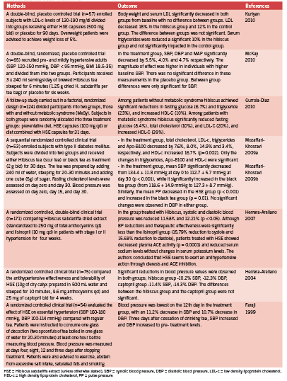
Human evidence We identified seven controlled human clinical trials evaluating hibiscus preparations. See Table 1. Five of the trials evaluated impact on blood pressure, three trials evaluated impact on serum lipid profiles, and one trial evaluated impact on plasma glucose levels. Only one trial failed to demonstrate a positive outcome; the trial evaluated impact to cholesterol levels and was compounded by recommendations to achieve reductions in body weight. Therefore both the hibiscus group and the control group experienced significant improvement in cholesterol profiles, with no significant difference between the two groups (Kuriyan 2010).
All five trials evaluating blood pressure demonstrated significant benefit, with a magnitude of effect ranging from 5-15% for reductions to systole and diastole (Faraji 1999, Herrera-Arellano 2007, Herrera-Arellano 2004, Mozaffari-Khosravi 2009b, McKay 2010). The two trials demonstrating positive impact to cholesterol levels revealed reductions in total cholesterol, LDL- cholesterol and triglyceride, as well as increased HDL-C levels (Gurrola-Diaz 2010, Mozaffari-Khosravi 2009a). The only trial to evaluate glucose levels likewise demonstrated significant improvement to glucose control (Gurrola- Diaz 2010).
Discussion Hibiscus, delivered in an array of dosage forms (standardized extracts, teas, decoctions), has demonstrated an impressive magnitude of benefit to key metabolic abnormalities, notably the constellation of abnormalities comprising the diagnostic criteria of the metabolic syndrome; glucose control, blood pressure, and plasma cholesterol levels, specifically triglyceride and HDL- cholesterol (as defined by Grundy 2004). The ease of delivery, the marginal cost of the medicine (especially in tea or decoction dosage forms), and an impressive safety profile make hibiscus a promising medicine for an array of very common clinical presentations. Longer- term trials are needed to determine if the effects of hibiscus are sustained beyond 30 days. Also, impact to blood glucose control deserves more research attention.
References:
Faraji MH, Tarkhani AHH. The effect of sour tea (Hibiscus sabdariffa) on essential hypertension. J Ethnopharmacol. 1999 Jun;65(3):231-6.
Frank T, Janben M, Netzel M, Strab G, Kler A, Kriesl E, Bitsch I. Pharmacokinetics of anthocyanidin-3-glycosides following consumption of Hibiscus sabdariffa L. extract. J Clin Pharmacol 2005 Feb;45(2):203-10.
Gurrola-Diaz CM, Garcia-Lopez PM, Sanchez-Enriquez S, Troyo-Sanroman R, Andrade-Gonzalex I, Gomez-Leyva JF. Effects of Hibiscus sabdariffa extract powder and prevention treatment (diet) on the lipid profiles of patients with metabolic syndrome (MeSy). Phytomedicine 2010 Jun;17(7):500-5.
Grundy SM, Brewer HB Jr, Cleeman JI, Smith SC Jr, Lenfant C. American Heart Association; National Heart, Lung, and Blood Institute. Definition of metabolic syndrome: Report of the National Heart, Lung, and Blood Institute/American Heart Association conference on scientific issues related to definition. Circulation. 2004 Jan 27;109(3):433-8.
Herrera-Arellano A, Flores-Romero S, Chavez-Soto MA, Tortoiello J. Effectiveness and tolerability of a standardized extract from Hibiscus sabdariffa in patients with mild to moderate hypertension: a controlled and randomized clinical trial. Phytomedicine 2004 Jul;11(5):375-82.
Herrera-Arellano A, Miranda-Sanchez J, Avila-Castro P, Herrera-Alvarez S, Jimenez-Ferrer JE, Zamilpa A, Roman-Ramos R, Ponce-Monter H, Tortoriello J. Clinical effects produced by a standardized herbal medicinal product of Hibiscus sabdariffa on patients with hypertension. A randomized, double-blind, lisinopril-controlled clinical trial. Planta Med 2007 Jan; 73(1): 6-12.
Huang CN, Chan KC, Lin WT, Su SL, Wang CJ, Peng CH. Hibiscus sabdariffa inhibits vascular smooth muscle cell proliferation and migration induced by high glucose – a mechanism involves connective tissue growth factor signals. J Agric Food Chem 2009 Apr 22;57(8):3073-9.
Kao ES, Tseng TH, Lee HJ, Chan KC, Wang CJ. Anthocyanin extracted from Hibiscus attenuate oxidized LDL-mediated foam cell formation involving regulation of CD36 gene. Chem Biol Interact 2009 May 15;179(2-3):212-8.
Kuriyan R, Kumar DR, R R, Kurpad AV. An evaluation of the hypolipidemic effect of an extract of Hibiscus Sabdariffa leaves in hyperlipidemic Indians: a double blind, placebo controlled trial. BMC Complement Altern Med. 2010 Jun 17;10:27.
McKay DL, Oliver Chen CY, Saltzman E, Blumberg JB. Hibiscus sabdariffa L. tea (Tisane) lowers blood pressure in prehypertensive and mildly hypertensive adults. J Nutr 2010 Feb;140(2):298-303.
Mozaffari-Khosravi H, Jalali-Khanabadi BA, Afkhami-Ardekani M, Fatehi F. Effects of sour tea (Hibiscus sabdariffa) on lipid profile and lipoproteins in patients with type II diabetes. J Altern Complement Med 2009a Aug;15(8):899-903.
Mozaffari-Khosravi H, Jalali-Khanabadi BA, Afkhami-Ardekani M, Fatehi F, Noori-Shadkam M. The effects of sour tea (Hibiscus sabdariffa) on hypertension in patients with type II diabetes. J Hum Hypertens 2009b Jan;23(1):48-54.
Nemeth K, Plumb GW, Berrin JG, Juge N, Jacob R, Naim HY, Williamson G, Swallow DM, Kroon PA. Deglycosylation by small intestinal epithelial cell beta-glucosidases is a critical step in the absorption and metabolism of dietary flavonoid glycosides in humans. Eur J Nutr. 2003 Jan;42(1):29-42.
Ojeda D, Jimenez-Ferrer E, Zamilpa A, Herrera-Arellano A, Tortoriello J, Alvarez L. Inhibition of angiotensin converting enzyme (ACE) activity by the anthocyanins delphinidin- and cyaniding-3-O-sambubiosides from Hibiscus sabdariffa. J Ethnopharmacol 2010 Jan 8;127(1):7-10.
Onyenekwe PC, Ajani EO, Ameh DA, Gamammiel KS. Antihypertensive effect of roselle (Hibiscus sabdariffa) calyx infusion in spontaneously hypertensive rats and a comparison of its toxicity with that in Wistar rats. Cell Biochem Func 1999 17: 199–206.
Rechner AR, Kuhnle G, Hu H, Roedig-Penman S, van den Braak MH, Moore KP, Rice-Evans CA. The metabolism of dietary polyphenols and the relevance to circulating levels of conjugated metabolites. Free Radic Res. 2002 Nov;36(11):1229-41.
Yang MY, Peng CH, Chan KC, Yang YS, Huang CN, Wang CJ. The hypolipidemic effect of Hibiscus sabdariffa polyphenols via inhibiting lipogenesis and promoting hepatic lipid clearance. J Agric Food Chem 2010 Jan 27;58(2):850-9.