Home Blog Page 261

Guiding Your Patients Along the Path to Good Mental Health

0

You’re undoubtedly noticing in your practice that as your patients get older, they’re also becoming more aware and concerned about declining mental ability.


We all know deep down inside that our short term memories are going to become less accessible and our cognitive pathways won’t be as crisp and efficient as they once were. And why should they be? After all, we have full lifetimes of memories to sort through and less urgency and need for efficiency in our daily lives. We’ve earned the right to relax, and yet we tend to hold ourselves to the standards by which we’ve led our lives. We allow these memory lapses and moments of absentmindedness to seem much more profound and portentous than they should.


It’s perfectly normal to experience phenomena like:


  1. Forgetting why we came into a room
  2. Having trouble remembering peoples names, even names we should know very well
  3. Being unable to come up with a noun to describe an object
  4. Getting confused or muddled when trying to recall familiar directions or processes


Some of your patients will come to you with a real sense of urgency if these lapses are becoming more frequent or making them noticeably less functional. And as a professional, you should be able to recognize signs and symptoms of phenomena like strokes and dementia and advise them about immediately seeking medical attention.


But when they come back to you, as so many of them will, because the medical establishment can’t provide satisfactory answers and solutions and your patients feel a need to be proactive – you need to be able to provide lifestyle advice that will help them manage and monitor their own mental health.


For many, that simply involves everyday things like maintaining a certain level of social engagement and physical activity, getting enough sleep and controlling mood and anxiety. Many of these things are at least partially manageable through diet. They should be seeking out natural source anti-oxidants from foods like walnuts and tomatoes and essential fatty acids through seafood or supplements. They would probably benefit from adding supplements to their regimen – even something as basic a good daily multivitamin to make sure they’re getting enough vitamin D, E, C and K.


And when they really want to get serious about feeding their brains – your patients will be heartened to know that you’re on top of all the trends and research about the very best steps and supplements they can take to look after their mental health.


While most people are very interested to learn about cutting edge anti-aging and dementia research – they are also aware of the lag time between scientific discoveries and the transformation of that new data into new treatments and medications.


We present a list of ingredients known to:

  • Increase their ability to concentrate and focus,
  • Help improve mental clarity,
  • Help prevent or alleviate memory loss,
  • Help maintain mental vitality and improve their capacity to assimilate new concepts,
  • Help deal with the depression that inevitably comes with aging and declining health.


Antioxidants inhibit oxidization and the resulting production of free radicals, which can damage or kill brain cells. Recommended anti-oxidants:


  • Acteyl-L-carnitine (ACL) A desirably bio-available amino acid that originates in our livers and kidneys, Acteyl-L-carnitine occurs naturally in the body and delivers great benefits to tissues such as the brain and heart. In addition to producing energy, it is shown to improve circulation in the brain. Although not recommended for people with under-active thyroid or a history of seizures, acetyl-L-carnitine can help boost energy production in the mitochondrial “power plants” in our cells, thereby increase mental energy, improve memory and protect the central nervous system.


  • Astaxanthin – a naturally occurring carotenoid found in marine life including fish, krill and crustaceans, astaxanthin is also found in bee resin and in the feathers of flamingos and storks. Perhaps the most potent of the xanthophyll carotenoids, astaxanthin is renowned for its anti-aging properties – and when it comes to brain health, it is known to decrease the risk of neurodegenerative disease by reducing oxidative stress.


  • Pycnogenol


Vitamin B 12:


High levels of the amino acid, homocysteine, contribute to brain shrinkage and increased risk of dementia. Vitamin B12 is an effective homocysteine suppressor.


As our society eats less meat – the main natural source of vitamin B12 – and gravitates toward more vegetarian diets, the risk of vitamin B12 deficiency increases. Our bodies don’t do a particularly good job of absorbing the nutrients we need and almost all vitamin B12 is absorbed through our small intestines. This process tends to get less efficient as we get older.


Traditional vitamin B supplements contain cyanocobalamine, in which the B12 is bound to cyanide. We depend on our livers to convert this to naturally occurring methylcobalamine – but of course, our livers also tend to do their job less well as we age. It makes sense to look beyond the traditional delivery system and

eliminate the need for conversion by taking methylcobalamine sublingual tablets (which dissolve under the tongue) and skin absorption (via a skin patch).


Omega-3s:


With almost 10 per cent of our brain’s matter comprised of the “essential fatty acids” known as omega-3s, it should come as no surprise that supplementing

Omega-3s may be the best way to prevent brain shrinkage and prevent nerve cell death. Recent studies even suggest that omega-3s can actually reverse some types of age related brain cell damage.


Efficient absorption becomes more of a challenge as we age – and since the shorter chained, plant based omega-3s known as alpha-linolenic acid (ALA) are harder for us to synthesize – we should probably lean toward the more readily usable, longer-chained, marine-based omega-3s docosahexanoic acid (DHA) and eicosapentanoic acid (EPA)


Phospholipids:

Because we each have different genetic backgrounds and all have different body chemistry (governed by diet, lifestyle and by the list of other medications we are taking), we all have different needs to restore balance and achieve and maintain optimal health. There are no one-size-fits-all solutions and it may be necessary to go through a process of figuring out what works best for each individual patient.


Nevertheless, most of the ingredients we present here are good for everyone – and many are essential in helping your maintain a healthy mind.

You & Your Patient: the Vital Bond

0

As in any other business, there is no single chartable route to success as a naturopath or an entrepreneur running your own clinic, but there are plenty of commonalities. The most essential part of the process of becoming established in a practice requires intense involvement in the community at large. The establishment of personal relationships with clients and the resulting referrals is often the single most important factor in determining the success of an ND’s practice.


Engagement with the Community


Dr. Brian Davies of Westcoast Integrative Health in North Vancouver stresses that it is important to “Leave your ego at the door. Clinical practice is 100% about supporting the patient.  Medicine is steeped in preconceptions, misconceptions, and beliefs about disease and treatment. As such, the focus needs to be on what the patient wants. Being a good listener and learning to manage expectations is one way I have worked to meet the needs of my patients.”


“I was extremely studious throughout my university career (University of Victoria and the Canadian College of Naturopathic Medicine.)  I wanted to know absolutely everything about medicine, to be able to recite it all forwards and backwards. Although my understanding of the discipline has been essential in clinical practice, knowledge isn’t everything. Equally important in my opinion, is how well you use this knowledge to educate and be educated by your patients.

“These concepts where not things that I learned in school but have allowed me to excel in my practice of naturopathic medicine. “


As far as actual marketing is concerns, Dr. Davies takes an even more low key approach. He volunteers in the community, teaches courses and gives lectures when the opportunity arises and says he gets 100 per cent of his business through referrals, a loyal patient base and networking in the community. He can identify a single occasion when he spent money on advertising during his ten plus years in practice.


Determining and Providing the Services that are Most Essential


Dr. Cathryn Coe, owner of North Vancouver’s Marine Drive Naturopathic Clinic has taken quite a different path. She regards it as vital to reach out to the people most likely to need your services – to educate them regarding possible causes of symptoms or conditions, and offer them solutions to these concerns. She is not averse to reaching outside of her immediate neighbourhood and putting the word out in nearby communities through advertising in both community newspapers, magazines and publications in the lower mainland. Even as an associate working out of someone else’s clinic, she advertised quite aggressively. Dr. Coe’s specialty is endocrinology and menopausal health and Marine Drive Naturopathic has benefitted and grown from her reputation and drive to get the word out about her services and personalized care. By establishing close and trusting relationships with her patients and offering her own expert insights and suggestions, the circle often widens – so that other family members come in to see her. This creates something of a RIPPLE effect, bringing attention and new clients to her associates – who themselves offer a diverse range of specialties. Her Marine Drive Naturopathic Clinic is experiencing considerable success and popularity, situated as it is in a well established community of people ranging from young professionals to active parents to productive and engaged retirees.


“I have used the same model both as an associate and as a business owner., “explains Dr. Coe, “identify the needs of your community, educate them about the steps that can be taken for them to overcome their symptoms or condition, and offer solutions. Even if patients don’t book with me directly, the type of advertising I do is educational, and for that reason benefits the profession as a whole.”


Every business decision Dr. Coe makes is calculated to deliver maximum benefit to her patients and her communities. She chose the physical location of her clinic because it is in the neighbourhood that has demonstrated the greatest demand for and appreciation of the work she does. “I can’t emphasize enough the importance of creating the right environment for your clients. In my clinic, your treatment begins right when you walk through the door.”


It’s All Personal


Regina’s Dr. Marika Geis agrees wholeheartedly with the need to establish personal relationships with patients.


Owner of Daziran Integrative Health, which operates out of the Cathedral Centre for Wellness in Regina, Dr. Geis enjoys a broad primary care practice emphasizing family medicine including women’s health issues, pediatric care, fertility counseling, perinatal care and stress syndromes (fatigue, insomnia, digestive issues, immune dysfunction and pain complexes).


Dr. Geis thinks of her patients almost as family – with whom she pursues relationships based on education and enlightenment: using nutritional counseling, botanical medicine, traditional Chinese medicine, clinical nutrition, lifestyle counseling and hydrotherapy – whatever speaks to and works for her individual patients. Determining and examining the physical, emotional, environmental and spiritual influences impacting the patient’s health – allows her to tailor treatments for each patient – bring them to a state of independence in their health, a sense of understand and having vital input into their own states of wellness.


Any entrepreneur in any field faces challenges common to the marketplace – weighing foot traffic against cachet, worrying about accessibility and signage, paying the rent, making budgets and managing cashflow.


But integrative healthcare practitioners have a unique role. Just by coming to see you in the first place, your patients are, as human beings, demonstrating their willingness – or even eagerness – to take responsibility for their own health, their own life and the lives of those they love.


As their caregiver, you can empower them further by giving them an understanding of their condition – and their options, roles and responsibilities in their own wellness.


Your toolbox contains an endless array of effective resources ranging from your understanding of physiological processes; knowledge of breaking research; awareness of and openmindedness to newly developed therapies and insights. But more than anything else – and perhaps more than in any other profession – your success depends on the relationships you create and maintain.


It’s not like you’re selling bread or auto parts or some other interchangeable product that your patients can find cheaper just down the road. The advice you give and the relationships you form are unique, valuable and irreplaceable. They come to you because of who you are and what you can give them that others can’t. The trust you build is your coin in trade.


Serene, Modern and Stylistically Unique

0

Just ten minutes from Halifax, in peaceful Upper Tantallon, rests a center of calm and healing known as Cornerstone Naturopathic – a state-of-the-art naturopathic clinic that represents the crossroads between a relaxing, regenerative environment and a sophisticated centre dedicated to the enhancement of medicine and continued recovery. Founder Dr. Ben Connolly believes that nature has an incredible power to help people heal and is an essential aspect of naturopathic medicine. As you arrive at Cornerstone Naturopathic and exit your vehicle you immediately hear a rushing river beside the clinic. While you take in your surroundings you see a beautiful view of St. Margaret’s Bay and a lush softwood forest. Finally you are greeted by a beautiful timber frame entrance to the clinic, this moment is when your healing begins. The new location for Cornerstone Naturopathic’s new clinic was chosen in Upper Tantallon outside of the city as almost all patients can benefit from a break away from the stresses of urban living.

Cornerstone-4-2

Due to the overarching principle of building a clinic in nature, there is little to no foot traffic on the street outside the clinic, yet always lots of patients inside. This has been made possible by word of mouth, many public speaking engagements in the community by their doctors, a strong social media presence, an excellent website and targeted trade shows.


Having just opened the doors to their new location in November 2014, Cornerstone is a one of a kind facility. With 7000 square feet, housing three full-time NDs, a full service infusion clinic, two registered massage therapists, a holistic nutritionist, a full gym onsite with personal training, an onsite nursery for patients to leave their children under supervision while they receive much needed treatment – it is one of the largest, most forward thinking Naturopathic Health Centers in Canada.


They are an evidenced based clinic, staying on top of the latest developments to help patients achieve an increased quality of life. This involves their doctors regularly travelling to conferences and taking courses across North America to bring back advanced treatments. In their state-of-the-art infusion room you will find patients receiving IV treatments such as tryptophan, taurine and aminosyn infusions for anxiety and depression through to L-carnitine and multi-vitamin infusions for mitochondria support for patients suffering from fatigue.

They also have an active full service multi-purpose/community education room which brings people into the clinic, which has featured smoothie workshops, movie presentations, birthing classes and make your own skin care product workshops.

Cornerstone-202


Each Naturopathic Doctor at Cornerstone has specific focuses in their practice. Dr. Colin MacLeod focuses on sports medicine and treating chronic and acute pain. He utilizes diet, lifestyle, acupuncture, injection and intravenous treatments for optimizing his patients’ health, performance and reducing their pain. Dr. Jillian Cole has a focus in mental and neurological health utilizing nutrition and supplements as well as individualized infusions. Dr. Ben Connolly divides his focuses between chronic and neurological pain utilizing nutrition and a variety of injection techniques including prolozone, neural and neuralprolo therapy. He also has a interested in cosmetic therapies.


Connolly believes that supporting the Naturopathic profession is as important as running a successful practice. Educating up and coming ND students outside of the classroom and providing them with opportunities to apply their knowledge hands on is crucial to a well-rounded ND upon graduation. They frequently have ND students in the clinic shadowing and currently run two externships a year with opportunity to increase up to six. They are also developing a residency program for new grads set to launch in 2016.

Cornerstone Naturopathic aims to bring Naturopathic Medicine to all of Nova Scotia and as a result have created a Cornerstone Satellite clinic in Yarmouth with three naturopathic doctors, while Dr. Colin MacLeod has created a satellite clinic serving the community in Port Hood, Cape Breton.


Resveratrol: A Review of Pre-clinical and Clinical Evidence in Cancer

0

Introduction

With the worldwide cancer incidence rate suspected to increase from 12.7 million cases per year in 2008, to a staggering 21.4 million cases per year in 2030, cancer prevention and treatment is crucial (Global Cancer Facts & Figures 2nd Edition, 2011). Recently, the use of natural dietary compounds as anti-cancer agents has become a large area of exploration in the field of cancer research. Accumulating evidence highlights the anti-carcinogenic and chemopreventive role of the phytolexin resveratrol, a compound found in the skin of grapes, red wine, chocolate, and certain berries (Kundu & Surh, 2008).

An initial study on the use of resveratrol as an anti-carcinogenic agent found that the topical application of resveratrol prevented tumor formation in mice (Jang et al., 1997). More recent preclinical data provides evidence for resveratrol’s protective properties against a multitude of cancers including colon, prostate, breast, hepatic, pancreatic, gastric, melanoma, and lung cancer (Carter, D’Orazio & Pearson, 2014).  At the molecular and cellular levels, resveratrol can act on DNA and proteins to inhibit the initiation, promotion, and progression stages of carcinogenesis (Jang et al., 1997; Devi, 2004).  Such chemopreventive effects can be achieved through resveratrol’s pro-apoptotic, anti-proliferative, anti-oxidant, anti-inflammatory, and anti-angiogenic properties (Carter, D’Orazio & Pearson, 2014; Boyce, Doehmer & Gooderham, 2004; Le Corre, Chalabi, Delort, Bignon & Bernard-Gallon, 2005).  In addition to its role as a chemopreventive agent, resveratrol has been shown to sensitize cancer cells to a number of chemotherapeutic agents, thus enhancing their chemotherapeutic potential (Fulda & Debatin, 2004; Santandreu, Valle, Oliver & Roca, 2011).

A concern for the implementation of resveratrol as part of cancer therapy is its bioavailability. Maintaining a clinically relevant dose is challenging as resveratrol is rapidly metabolized in the body (Walle et al., 2004).  However, recent research has analyzed different ways to improve resveratrol’s bioavailability in the human body (Howells et al., 2011; Dellinger, Garcia &  Meyskens, 2014; la Porte et al., 2010).  The objective of this paper is to conduct a review of the pre-clinical and clinical evidence surrounding the use of resveratrol as a chemopreventive agent in cancer, its bioavailability in humans, and its ability to sensitize cancer cells to conventional chemotherapeutics.


Chemopreventive Properties

Apoptosis & Cell Cycle Arrest

Human cell line studies have highlighted the role of resveratrol as a pro-apoptotic and anti-proliferative phytochemical against a variety of cancers including colon, pancreatic, breast, lung, prostate, gastric, and skin cancer cell lines (Temraz, Mukherji & Shamseddine, 2013; Shamim et al., 2012; Shi et al., 2011; Luo, Yang, Schulte, Wargovich & Wang, 2013; Kai, Samuel & Levenson, 2010; Chung, Lim & Lee, 2013; Kim, 2012).  Such studies have focused on understanding resveratrol’s underlying pro-apoptotic and anti-proliferative mechanisms, with most activity surrounding interactions with DNA and proteins, including the activation of tumour suppressor protein p53, activation of cell cycle inhibitors p21 and p27, and the up-regulation of pro-apoptotic proteins such as Bax (Malhotra, Nair & Dhawan, 2012).  Moreover, resveratrol has been found to inhibit major pathways involved in cell growth and migration including the WNT, MAPK-ERK, and PI3K-AKT signalling pathways (Refer to Table 1 for further details) (Azmi et al., 2013; Gescher, Steward & Brown, 2013; Carter, D’Orazio & Pearson, 2014).

Similarly, in vivo studies using lung, skin, and pancreatic mouse models demonstrate that resveratrol administration leads to a reduction in tumour growth (Malhotra, Nair & Dhawan, 2012; Gescher, Steward & Brown, 2013; Carter, D’Orazio & Pearson, 2014).  Although one study found resveratrol to have pro-apoptotic effects on a normal pancreatic cell line, the study failed to provide data for this observation, or elaborate on the extent of cytotoxicity (Azmi et al., 2013).  Multiple other studies suggest that resveratrol preferentially targets cancer cells, while simultaneously protecting normal host cells from unwanted growth arrest and apoptosis (Luo, Yang, Schulte, Wargovich & Wang, 2013; Rusin, Zajkowicz & Butkiewicz, 2009; Tyagi et al., 2011).


Antioxidant

Low dose resveratrol (<50μM) has been shown to promote antioxidant properties by regulating the generation of reactive oxygen species, while high doses are suggested to promote pro-oxidant activity (Shrotriya, Agarwal & Sclafani, 2015; Luo, Yang, Schulte, Wargovich & Wang, 2013). Studies using cell line and animal models of skin, colon, lung, and hepatic cancers propose that resveratrol enhances the activity of the transcription factor Nrf-2, which stimulates the expression of antioxidant enzymes such as glutathione S-transferase, catalase, glutathione peroxidase, and superoxide dismutase (Refer to Table 2 for further details) (Shrotriya, Agarwal & Sclafani, 2015; Chiou et al., 2011; Tan et al., 2012; Khan et al., 2013; Bishayee, Barnes, Bhatia, Darvesh & Carroll, 2010). Hence, this is important given that accumulating evidence proposes an association of various cancers with low activities of antioxidant enzymes (Arsova-Sarafinovska et al., 2009; Sharma, Tripathi, Satyam & Kumar, 2009).


Anti-inflammatory & Anti-angiogenesis Properties

Resveratrol has also been shown to have anti-inflammatory and anti-angiogenic properties. Since tumor cells utilize the body’s inflammatory response mechanisms to induce angiogenesis for survival, reduced inflammatory responses may help induce tumor cell death by preventing angiogenesis and cell proliferation (Dalgleish & Haefner, 2006; Bishayee, Barnes, Bhatia, Darvesh & Carroll, 2010). Resveratrol has been shown to down-regulate the expression of the pro-inflammatory enzyme COX-2 in a hepatic cancer rat model, and reduce the generation of the inflammatory mediator nitric oxide in colon cancer cell lines (Bishayee, Barnes, Bhatia, Darvesh & Carroll, 2010; Panaro, Carofiglio, Acquafredda, Cavallo & Cianciulli, 2012). Additionally, a study found that resveratrol exerts anti-angiogenic effects in a melanoma cell line by decreasing levels of vascular endothelial growth factor (Trapp, Parmakhtiar, Papazian, Willmott & Fruehauf, 2010). In humans with prostate cancer, resveratrol inhibits the activities of metastasis-associated antigen leading to p53 activation, as well as reduced invasion and angiogenesis (Refer to Table 3 for further details) (Levenson, Kumar & Zhang, 2014).


Prevention of Cancer Onset

In addition to the protective properties that resveratrol has against existing cancers, it can also play a role in preventing cancer onset. Cancer prevention often involves inhibition of cytochrome P450 (CYP) enzymes that can activate environmental carcinogens. For example, a group of CYP enzymes metabolize alcohol into acetaldehyde, which then interacts with DNA and increases the risk of head and neck cancers (Chow et al., 2010; Shrotriya, Agarwal & Sclafani, 2015). In lung, breast, and colon cancer cell lines, resveratrol was found to reduce CYP1A1 mRNA accumulation by approximately 50%, thus reducing the amount of enzyme available to activate carcinogens (Perdew et al., 2010). Moreover, CYP enzyme activity was measured in a clinical study consisting of 42 healthy volunteers taking 1g of resveratrol daily for four weeks. Their results suggest that pharmacologic doses of resveratrol have the ability to upregulate or down-regulate CYP enzyme levels. This can interfere with the metabolism of cholesterol lowering statins and chemotherapeutics, thus necessitating caution before administering resveratrol in clinical settings (Refer to Table 4 for further details) (Chow et al., 2010).


Bioavailability

Due to resveratrol’s rapid metabolism, a challenge for clinical implementation is the difficulty in maintaining a therapeutically relevant level of resveratrol in the blood (Singh, Ndiaye & Ahmad, 2014).  Bioavailability studies have shown that trans-resveratrol, the biologically active form, is quickly absorbed into the circulation, with maximal plasma concentrations achieved after 30 minutes (Bishayee, 2009).  However, phase II metabolism in the liver rapidly catabolizes resveratrol into glucuronide and sulfate conjugates (Walle et al., 2004).  Consequently, serum analysis 4 hours after the administration of a dietary-relevant dose of resveratrol at 25mg/70kg reveals that only 2% of total resveratrol remains in its unmodified form, indicating low oral bioavailability (Goldberg et al. 2003).  Moreover, in a recent clinical trial, where a high dose of resveratrol (5g) was administered to 10 healthy participants for 29 days, plasma resveratrol concentrations were measured to be approximately 4μM.  This concentration is below levels previously shown to have chemopreventive effects in preclinical studies (Brown et al., 2010; Boocock et al., 2007).

Despite its inherently low blood concentration, resveratrol can be rapidly absorbed to exert chemopreventive effects (Walle et al., 2004). Urinary excretion data determined that more than 70% of a single 100mg dose of resveratrol was absorbed into cells, suggesting that intratissular and intracellular levels may be higher than those measured in the plasma  (Walle et al., 2004; Patel et al., 2013). Moreover, recent evidence suggests that certain resveratrol conjugates can be enzymatically returned to their trans-resveratrol form, or may even promote anti-proliferative effects on their own (Zhu et al., 2012).

Modifications to resveratrol have been made to address the difficulties surrounding its bioavailability and rapid metabolism. For example, with a single dose of SRT501, a micronized oral form of resveratrol, mean plasma levels were 3.6 times higher than a similar dose of non-micronized resveratrol (Howells et al., 2011).  It may also be useful to modify the molecular structure of resveratrol.  Pterostilbene, a dimethylated analogue of resveratrol, is metabolized slower, thus contributing to improved bioavailability (Dellinger, Garcia &  Meyskens, 2014).  In addition, a study examining the pharmacokinetics of resveratrol revealed that when taken with a high fat meal, the maximal plasma concentration of resveratrol was reduced (la Porte et al., 2010). Therefore, through micronization, development of resveratrol analogues, or avoidance of a high fat diet, it may be possible to address resveratrol’s bioavailability concerns.


Clinical Trials

All clinical trials to date are conveniently summarized in Table 5 of the Appendix. From the limited number of clinical trials, there appears to be conflicting results; however, the positive results justify looking further into resveratrol as a potential chemopreventive agent.

A randomized controlled trial of participants with colorectal cancer and hepatic metastasis (n=9) found that receiving 5g SRT501 for 14 days resulted in a 39% increase in cleaved caspase 3.  The study also analyzed other common markers of apoptosis, but found no significant difference in levels between the two groups. While this evidence is not conclusive, it does suggest that resveratrol may promote apoptosis in humans (Howells et al., 2011).

Another study of patients with colorectal cancer (n=20) presented positive evidence for resveratrol as a chemopreventive agent. In this study, daily doses of 0.5g or 1.0g were administered over eight days to participants prior to surgical resection. A comparison of pre-intervention and post-intervention tissue samples demonstrated a significant 5% reduction in Ki-67 staining, a marker of tumour cell proliferation. Despite the small dose, high enough resveratrol concentrations were achieved within the gastrointestinal tract to produce anti-carcinogenic effects (Patel et al., 2010). Due to the small study populations and high variability between participants in existing clinical trials, it is difficult to make clear clinical recommendations on the effectiveness of resveratrol as a cancer preventive or therapeutic agent.

Some evidence suggests that resveratrol may have harmful adverse effects.  One study consisting of participants with relapsed or refractory multiple myeloma (MM) (n=24) found that doses of 5.0g SRT501 combined with bortezomib produced serious adverse effects.  During the study, five serious adverse events of renal toxicity were observed in addition to two deaths, one potentially related to the treatment, causing researchers to end the study early  (Popat et al., 2013).  Results this harmful have not been found in other studies using a 5g dose of SRT501; Howells et al. found mild gastrointestinal adverse effects reported by participants. As well, in a repeat dose study with healthy volunteers (n=40), only mild short-term gastrointestinal adverse effects were noted in participants taking 2.5g or 5g doses of resveratrol for 29 days (Brown et al., 2010).  It has been reported that about 50% of patients with MM develop renal impairment as the disease progresses. Additionally, three out of the five participants who experienced renal toxicity as an adverse event began the study with elevated blood creatinine levels, potentially indicating the presence of pre-existing renal dysfunction (Popat et al., 2013). Thus, participants in the Popat et al. study may have been at a higher baseline risk of developing renal failure.  Nonetheless, as a precaution, we propose that resveratrol should not be used in those with pre-existing kidney conditions. Despite the unexpected finding of renal toxicity in MM patients taking SRT501, these results do not indicate that SRT501 or resveratrol are unsafe for other cancer populations. This, however, does require further research into the safety and dosing of resveratrol.


Chemosensitization of Resveratrol

Based on evidence from clinical trials to date, resveratrol may not be sufficiently efficacious to be considered as an alternative to conventional cancer therapy; however, it does have the potential to be integrated into existing cancer treatment regimens. Accumulating evidence highlights resveratrol’s role as a chemosensitizing agent to a variety of chemotherapeutic drugs. In vitro studies with melphalan, for instance, have demonstrated that resveratrol can sensitize human breast cancer cells to melphalan-induced apoptosis, S-phase cell cycle arrest, and caspase activation (Casanova et al., 2012). Additionally, resveratrol was found to enhance the effects of cyclophosphamide (CPA) in human breast cancer cell lines; thus, a lower CPA dosage is required to achieve therapeutic effects. This minimizes CPA-associated chemotoxicity and maximizes anti-cancer effects (Singh et al., 2011).

Similarly, in vivo studies demonstrated resveratrol’s ability to decrease the required dose of cisplatin, gefitinib and paclitaxel in mice implanted with human lung cancer cells (Zhao, Bao, Qi & You, 2010). In a murine model of human pancreatic cancer, resveratrol was shown to sensitize pancreatic cancer cells and potentiate the effects of gemcitabine through the inhibition of NF-kB activation and expression of cell survival proteins (Harikumar et al., 2010).

Various chemotherapeutic agents exert pro-oxidant activity, but despite resveratrol’s anti-oxidant properties, no antagonistic effects were observed in the aforementioned studies.  Some studies, however, reported the potential for resveratrol to suppress apoptosis induced by daunorubicin and two mitotic inhibitors, paclitaxel and vincristine (Gupta, Kannappan, Reuter, Kim & Aggarwal, 2011).

Resveratrol’s chemosensitization effects may also be dependent on dosage; in vitro studies involving human colon cancer cells demonstrated that resveratrol synergistically promoted 5-fluorouracil (5-FU)-mediated apoptosis at high doses of both resveratrol (200μM) and 5-FU (120μM), as well as with lower doses of both resveratrol (15μM) and 5-FU (0.5μM) (Chan, Phoo, Clement, Pervaiz & Lee, 2008; Mohapatra et al., 2011). Although resveratrol has the potential to be used in combination with various chemotherapeutic agents, further studies are needed before this combination therapy could be implemented in clinical settings.


Discussion

This review summarizes current evidence on resveratrol as a factor with pro-apoptotic, anti-proliferative, anti-inflammatory, anti-angiogenic, antioxidant and chemosensitizing properties. Cell line and animal studies have highlighted this phytochemical’s efficacy against a variety of cancers including colon, pancreatic, breast, lung, prostate, gastric, hepatic and skin cancer. Due to its many modes of anti-cancer activity, from changes in DNA expression to protein interactions and pathway-modulatory activity, resveratrol has garnered significant attention in the field of cancer research.

Although cell line and animal studies have suggested resveratrol to be efficacious against cancer cells and tumours, clinical data on resveratrol’s chemopreventive potential in humans is conflicting. Small samples and unstandardized populations across clinical trials, in addition to evidence suggesting potential adverse effects of resveratrol, highlight the need for further human research.  Thus, the current research on resveratrol does not permit a clinical recommendation to be made, except that as a precautionary measure, it should not be used in patients with pre-existing renal conditions (Popat et al., 2013).

Future research should focus on evaluating the effectiveness and safety of resveratrol in a variety of different cancer types. Moreover, a look into resveratrol analogues with significantly more potency and improved bioavailability, such as pterostilbene, are also recommended.  Pterostilbene has been shown to have more potent inhibitory effects on the growth of three colon cancer cell lines, as measured by levels of cleaved caspase-3 (Nutakul et al., 2011; Dellinger, Garcia &  Meyskens, 2014).  Ultimately, in clinical practice, resveratrol will likely be used as an adjuvant therapy; therefore, beyond researching resveratrol as a monotherapeutic agent, future studies should further investigate resveratrol in combination with classic chemotherapeutic agents. In addition, since resveratrol displayed antagonistic effects with mitotic inhibitors such as paclitaxel and vincristine, we recommend further exploring the interaction of resveratrol with mitotic inhibitors (Gupta, Kannappan, Reuter, Kim & Aggarwal, 2011).


Conclusion

Additional studies are needed to further investigate and understand resveratrol’s chemopreventive effects when combined with chemotherapeutic agents.  Nonetheless, it is evident that the difficulties surrounding resveratrol’s bioavailability can be addressed and that there are potential benefits for implementing resveratrol as a combinatory therapy in clinical settings in the future.

References

Arsova-Sarafinovska, Z., Eken, A., Matevska, N., Erdem, O., Sayal, A., & Savaser, A., Banev, S., Petrovski, D., Dzikova, S., Georgiev, V., Sikole, A., Ozgok, Y., Suturkova, L., Dimovski, A., & Aydin, A. (2009). Increased oxidative/nitrosative stress and decreased antioxidant enzyme activities in prostate cancer.Clinical Biochemistry, 42(12), 1228-1235.


Azmi, M. N., Din, M. F. M., Kee, C. H., Suhaimi, M., Ping, A. K., Ahmad, K., Nafiah, M.A., Thomas, N.F., Mohamad, K., Hoong, L.K., & Awang, K. (2013). Design, synthesis and cytotoxic evaluation of o-carboxamido stilbene analogues. International journal of molecular sciences, 14(12), 23369-23389.


Bishayee, A. (2009). Cancer prevention and treatment with resveratrol: from rodent studies to clinical trials. Cancer prevention research, 2(5), 409-418.


Bishayee, A., Barnes, K., Bhatia, D., Darvesh, A., & Carroll, R. (2010). Resveratrol Suppresses Oxidative Stress and Inflammatory Response in Diethylnitrosamine-Initiated Rat Hepatocarcinogenesis. Cancer Prevention Research, 3(6), 753-763.


Bishayee A, Waghray A, Barnes K, Mbimba T, Bhatia D, Chatterjee M & Darvesh A. (2010). Suppression of the Inflammatory Cascade is Implicated in Resveratrol Chemoprevention of Experimental Hepatocarcinogenesis. Pharmaceutical Research, 27(6), 1080-1091.


Boocock, D. J., Faust, G. E., Patel, K. R., Schinas, A. M., Brown, V. A., Ducharme, M. P., Booth, T. D., Crowell, J. A, Perloff, M., Gescher, A. J., Steward, W. P., & Brenner, D. E. (2007). Phase I dose escalation pharmacokinetic study in healthy volunteers of resveratrol, a potential cancer chemopreventive agent. Cancer Epidemiology Biomarkers & Prevention, 16(6), 1246-1252.


Boyce, A., Doehmer, J., & Gooderham, N. J. (2004). Phytoalexin resveratrol attenuates the mutagenicity of the heterocyclic amines 2-amino-1-methyl-6-phenylimidazo [4, 5-b] pyridine and 2-amino-3, 8-dimethylimidazo [4, 5-f] quinoxaline. Journal of Chromatography B, 802(1), 217-223.


Brown, V. A., Patel, K. R., Viskaduraki, M., Crowell, J. A., Perloff, M., Booth, T. D., Vasilinin, G., Sen, A., Schinas, A. , Piccirilli, G., Brown, K., Steward, W. P., Gescher, A. J., & Brenner, D. E. (2010). Repeat dose study of the cancer chemopreventive agent resveratrol in healthy volunteers: safety, pharmacokinetics, and effect on the insulin-like growth factor axis. Cancer research, 70(22), 9003-9011.

Carter, L. G., D’Orazio, J. A., & Pearson, K. J. (2014). Resveratrol and cancer: focus on in vivo evidence. Endocrine-related cancer, 21(3), R209-R225.


Casanova, F., Quarti, J., da Costa, D. C. F., Ramos, C. A., da Silva, J. L., & Fialho, E. (2012). Resveratrol chemosensitizes breast cancer cells to Melphalan by cell cycle arrest. Journal of Cellular Biochemistry, 113(8), 2586–2596.


Chan, J. Y., Phoo, M. S., Clement, M.-V., Pervaiz, S., & Lee, S. C. (2008). Resveratrol displays

converse dose-related effects on 5-fluorouracil-evoked colon cancer cell apoptosis – The roles of

caspase-6 and p53. Cancer Biology & Therapy, 7(8), 1305–1312.


Chiou, Y., Tsai, M., Nagabhushanam, K., Wang, Y., Wu, C., Ho, C., & Pan, M. (2011). Pterostilbene Is More Potent than Resveratrol in Preventing Azoxymethane (AOM)-Induced Colon Tumorigenesis via Activation of the NF-E2-Related Factor 2 (Nrf2)-Mediated Antioxidant Signaling Pathway. Journal Of Agricultural And Food Chemistry, 59(6), 2725-2733.


Chow, H., Garland, L., Hsu, C., Vining, D., Chew, W., Miller, J., Perloff, M., Crowell, J. A., & Alberts, D. S. (2010). Resveratrol Modulates Drug- and Carcinogen-Metabolizing Enzymes in a Healthy Volunteer Study. Cancer Prevention Research, 3(9), 1168-1175.


Chung, M. Y., Lim, T. G., & Lee, K. W. (2013). Molecular mechanisms of chemopreventive phytochemicals against gastroenterological cancer development. World journal of gastroenterology: WJG, 19(7), 984.


Dalgleish, A., & Haefner, B. (2006). The link between inflammation and cancer (pp. 12-14). New York: Springer.


Dellinger, R. W., Garcia, A. M. G., & Meyskens, Jr, F. L. (2014). Differences in the glucuronidation of resveratrol and pterostilbene: Altered enzyme specificity and potential gender differences. Drug metabolism and pharmacokinetics, 29(2), 112-119.


Devi, P. U. (2004). Basics of carcinogenesis. Health Adm, 17(1), 16-24.


Fulda, S., & Debatin, K.-M. (2004). Sensitization for anticancer drug-induced apoptosis by the chemopreventive agent resveratrol. Oncogene, 23(40), 6702–6711.


Gescher, A., Steward, W. P., & Brown, K. (2013). Resveratrol in the management of human cancer: how strong is the clinical evidence?. Annals of the New York Academy of Sciences, 1290(1), 12-20.


Global Cancer Facts & Figures. (2011) (2nd ed., p. 1). Atlanta. Retrieved from http://www.cancer.org/acs/groups/content/@epidemiologysurveilance/documents/document/acspc-027766.pdf


Goldberg, D. M., Yan, J., & Soleas, G. J. (2003). Absorption of three wine-related polyphenols in three different matrices by healthy subjects. Clinical biochemistry, 36(1), 79-87.


Gupta, S. C., Kannappan, R., Reuter, S., Kim, J. H., & Aggarwal, B. B. (2011). Chemosensitization of tumors by resveratrol. Annals of the New York Academy of Sciences, 1215(1), 150–160.


Harikumar, K. B., Kunnumakkara, A. B., Sethi, G., Diagaradjane, P., Anand, P., Pandey, M. K., Gelovani, J., Krishnan, S., Guha, S., & Aggarwal, B. (2010). Resveratrol, a multitargeted agent, can enhance antitumor activity of Gemcitabine in vitro and in orthotopic mouse model of human pancreatic cancer. International Journal of Cancer, 127(2), 257–268.


Howells, L. M., Berry, D. P., Elliott, P. J., Jacobson, E. W., Hoffmann, E., Hegarty, B., Brown, K., Steward, W. P., & Gescher, A. J. (2011). Phase I randomized, double-blind pilot study of micronized resveratrol (SRT501) in patients with hepatic metastases—safety, pharmacokinetics, and pharmacodynamics. Cancer Prevention Research, 4(9), 1419-1425.

Jang, M., Cai, L., Udeani, G. O., Slowing, K. V., Thomas, C. F., Beecher, C. W., Fong H.H., Farnsworth, N. R., Kinghorn A. D., Mehta R. G., Moon R. C., & Pezzuto, J. M. (1997). Cancer chemopreventive activity of resveratrol, a natural product derived from grapes. Science, 275(5297), 218-220.


Kai, L., Samuel, S. K., & Levenson, A. S. (2010). Resveratrol enhances p53 acetylation and apoptosis in prostate cancer by inhibiting MTA1/NuRD complex.International Journal of Cancer, 126(7), 1538-1548.


Khan, M., Chen, H., Wan, X., Tania, M., Xu, A., Chen, F., & Zhang, D. (2013). Regulatory effects of resveratrol on antioxidant enzymes: A mechanism of growth inhibition and apoptosis induction in cancer cells. Molecules And Cells, 35(3), 219-225.


Kim, M. Y. (2012). Nitric oxide triggers apoptosis in A375 human melanoma cells treated with capsaicin and resveratrol. Molecular medicine reports, 5(2), 585-591.


Kundu, J. K., & Surh, Y. J. (2008). Cancer chemopreventive and therapeutic potential of resveratrol: mechanistic perspectives. Cancer letters, 269(2), 243-261.


la Porte, C., Voduc, N., Zhang, G., Seguin, I., Tardiff, D., Singhal, N., & Cameron, D. W. (2010). Steady-state pharmacokinetics and tolerability of trans-resveratrol 2000mg twice daily with food, quercetin and alcohol (ethanol) in healthy human subjects. Clinical pharmacokinetics, 49(7), 449-454.


Le Corre, L., Chalabi, N., Delort, L., Bignon, Y. J., & Bernard‐Gallon, D. J. (2005). Resveratrol and breast cancer chemoprevention: molecular mechanisms. Molecular nutrition & food research, 49(5), 462-471.


Levenson A, Kumar A, Zhang X. (2014). MTA family of proteins in prostate cancer: biology, significance, and therapeutic opportunities. Cancer Metastasis Rev., 33(4), 929-942.


Luo, H., Yang, A., Schulte, B. A., Wargovich, M. J., & Wang, G. Y. (2013). Resveratrol induces premature senescence in lung cancer cells via ROS-mediated DNA damage. PloS one, 8(3), e60065.


Malhotra, A., Nair, P., & Dhawan, D. K. (2012). Curcumin and resveratrol in combination modulates benzo (a) pyrene-induced genotoxicity during lung carcinogenesis. Human & experimental toxicology, 31(12), 1199-1206.


Nutakul, W., Sobers, H. S., Qiu, P., Dong, P., Decker, E. A., McClements, D. J., & Xiao, H. (2011). Inhibitory effects of resveratrol and pterostilbene on human colon cancer cells: a side-by-side comparison. Journal of agricultural and food chemistry, 59(20), 10964-10970.


Panaro M, Carofiglio V, Acquafredda A, Cavallo P, Cianciulli A. (2012). Anti-inflammatory effects of resveratrol occur via inhibition of lipopolysaccharide-induced NF-κB activation in Caco-2 and SW480 human colon cancer cells. British Journal of Nutrition, 108(09), 1623-1632.


Patel, K. R., Andreadi, C., Britton, R. G., Horner-Glister, E., Karmokar, A., Sale, S., Brown, V. A., Brenner, D. E., Singh, R., Steward, W. P., Gescher, A. J., & Brown, K. (2013). Sulfate metabolites provide an intracellular pool for resveratrol generation and induce autophagy with senescence. Science translational medicine, 5(205), 205ra133-205ra133.

Patel, K. R., Brown, V. A., Jones, D. J., Britton, R. G., Hemingway, D., Miller, A. S., West, K. P, Booth, T. D., Perloff, M., Crowell, J. A., Brenner, D. E., Steward, W. P., Gescher, A. J., &

Brown, K. (2010). Clinical pharmacology of resveratrol and its metabolites in colorectal cancer patients. Cancer research, 70(19), 7392-7399.


Perdew, G., Hollingshead, B., DiNatale, B., Morales, J., Labrecque, M., Takhar, M. Tam, K. J., & Beischlag, T.V., (2010). Estrogen Receptor Expression Is Required for Low-Dose Resveratrol-Mediated Repression of Aryl Hydrocarbon Receptor Activity. Journal Of Pharmacology And Experimental Therapeutics, 335(2), 273-283.


Popat, R., Plesner, T., Davies, F., Cook, G., Cook, M., Elliott, P.,Jacobson, E., Gumbleton, T., Oakervee, H., Cavenagh, J., & Cavenagh, J. (2013). A phase 2 study of SRT501 (resveratrol) with bortezomib for patients with relapsed and or refractory multiple myeloma. British journal of haematology, 160(5), 714-717.


Rusin, M., Zajkowicz, A., & Butkiewicz, D. (2009). Resveratrol induces senescence-like growth inhibition of U-2 OS cells associated with the instability of telomeric DNA and upregulation of BRCA1. Mechanisms of ageing and development, 130(8), 528-537.


Santandreu, F. M., Valle, A., Oliver, J., & Roca, P. (2011). Resveratrol Potentiates the Cytotoxic Oxidative Stress Induced by Chemotherapy in Human Colon Cancer Cells. Cellular Physiology and Biochemistry, 28(2), 219–228.


Shamim, U., Hanif, S., Albanyan, A., Beck, F. W., Bao, B., Wang, Z., Banerjee, S., Sarkar, F. H., Mohammad, R. M., Hadi, S. M., & Azmi, A. S. (2012). Resveratrol‐induced apoptosis is enhanced in low pH environments associated with cancer. Journal of cellular physiology, 227(4), 1493-1500.


Sharma, A., Tripathi, M., Satyam, A., & Kumar, L. (2009). Study of antioxidant levels in patients with multiple myeloma. Leukemia & Lymphoma, 50(5), 809-815.

Shi, Y., Yang, S., Troup, S., Lu, X., Callaghan, S., Park, D. S., Xing, Y., & Yang, X. (2011). Resveratrol induces apoptosis in breast cancer cells by E2F1-mediated up-regulation of ASPP1. Oncology reports, 25(6), 1713-1719.


Shrotriya, S., Agarwal, R., & Sclafani, R. A. (2015). A Perspective on Chemoprevention by Resveratrol in Head and Neck Squamous Cell Carcinoma. In Biological Basis of Alcohol-Induced Cancer (pp. 333-348). Springer International Publishing.


Singh, C. K., Ndiaye, M. A., & Ahmad, N. (2014). Resveratrol and cancer: challenges for clinical translation. Biochimica et Biophysica Acta (BBA)-Molecular Basis of Disease.


Singh, N., Nigam, M., Ranjan, V., Zaidi, D., Garg, V. K., Sharma, S., Rashmi, C., Shankar, R., Kumar, S., Sharma R., Mitra, K., Balapure, A.K., Rath, S. (2011). Resveratrol as an adjunct therapy in cyclophosphamide-treated MCF-7 cells and breast tumor explants. Cancer Science, 102(5), 1059–1067.


Tan, X., Marquardt, G., Massimi, A., Shi, M., Han, W., & Spivack, S. (2012). High-Throughput Library Screening Identifies Two Novel NQO1 Inducers in Human Lung Cells. Am J Respir Cellular and Molecular Biology, 46(3), 365-371.


Temraz, S., Mukherji, D., & Shamseddine, A. (2013). Potential targets for colorectal cancer prevention. International journal of molecular sciences, 14(9), 17279-17303.


Trapp V, Parmakhtiar B, Papazian V, Willmott L, Fruehauf J. (2010). Anti-angiogenic effects of resveratrol mediated by decreased VEGF and increased TSP1 expression in melanoma-endothelial cell co-culture. Angiogenesis, 13(4), 305-315.


Tyagi, A., Gu, M., Takahata, T., Frederick, B., Agarwal, C., Siriwardana, S., Agarwal, R., & Sclafani, R. A. (2011). Resveratrol selectively induces DNA damage, independent of Smad4 expression, in its efficacy against human head and neck squamous cell carcinoma. Clinical Cancer Research, 17(16), 5402-5411.


Walle T, Hsieh F, DeLegge MH, Oatis JE Jr, Walle UK.(2004). High absorption but very low bioavailability of oral resveratrol in humans. Drug Metabolism and Disposition, 32(12):1377-1382.

Zhao, W., Bao, P., Qi, H., & You, H. (2010). Resveratrol down-regulates survivin and induces

apoptosis in human multidrug-resistant SPC-A-1/CDDP cells. Oncology Reports, 23(1),

279–286.

Zhu, W., Qin, W., Zhang, K., Rottinghaus, G. E., Chen, Y. C., Kliethermes, B., & Sauter, E. R. (2012). Trans-resveratrol alters mammary promoter hypermethylation in women at increased risk for breast cancer. Nutrition and cancer, 64(3), 393-4

Curcumin as an adjuvant therapy to conventional cancer treatments

0

Introduction

Forty percent of Canadians develop cancer in their lifetime, and twenty-five percent of these cases are fatal (Canadian Cancer Society 2014). The standards of care for cancer are chemotherapy, radiation, and/or surgery, because these modalities have demonstrated the highest treatment efficacy in clinical practice (Canadian Cancer Society 2012, Canadian Cancer Society 2015). However, patients diagnosed with chemotherapy- and radiotherapy-resistant cancers face treatment challenges (Li 2014). Thus, treatments adjuvant to conventional therapy are being explored.

Turmeric (Curcuma longa) is a plant native to Asia (Shishodia 2005). This nutraceutical has garnered interest as a complementary cancer therapy, as observational evidence has demonstrated low rates of colorectal, prostate, and lung cancers in Asia, where high amounts of turmeric are consumed (Aggarwal 2003, Lao 2006, Sinha 2003).

Curcumin (diferuloylmethane) is the most thoroughly researched active component of turmeric (Chattopadhyay 2004, Lao 2006, Shishodia 2005). Research evidence demonstrates that curcumin is more effective when used in conjunction with chemotherapy than as a stand-alone therapy (Kusuhara 2012, Lin 2007). This is due to its ability to downregulate resistance proteins (Guo 2014, Rana 2015, Roy 2014) and modulate cancer stem cells, which are both integral mechanisms to cancer resistance, metastasis, and recurrence (Buhrmann 2014, Shakibaei 2014). Moreover, curcumin antagonizes many of chemotherapy’s negative side effects such as promotion of cell proliferation through NF-kB (Cabrespine-Faugeras 2010, Melisi 2007). This paper investigates the clinical potential for curcumin use with conventional therapy to improve treatment outcomes in a variety of cancers.


General Cancer Pathways

Figure 1: Cellular pathways involved in cancer pathology and the effects of curcumin and chemotherapy

CHART

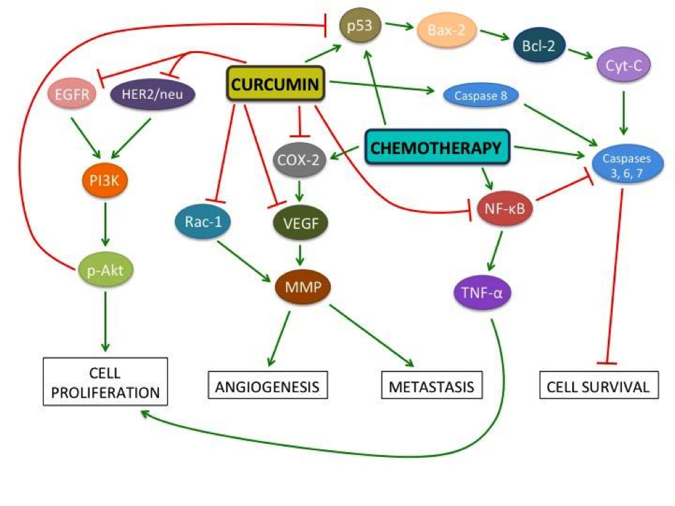
This diagram illustrates the effects of curcumin and chemotherapy on various molecules involved in cancer. Curcumin decreases angiogenesis, tumour cell proliferation, metastasis, and cancer cell survival. Nuclear factor-kappaB (NF-kB) activation facilitates tumour cell survival by downregulating caspase-3, -6, and -7, which are factors integral to cellular apoptosis (Cabrespine-Faugeras 2010, Melisi 2007). Paradoxically, chemotherapeutic drugs often upregulate NF-kB (Kamat 2009, Tharakan 2010), while curcumin inhibits it (Cabrespine-Faugeras 2010). NF-kB upregulates the inflammatory factor TNF-, which leads to increased cellular proliferation. Curcumin and chemotherapy drugs inhibit Bcl-2 – an apoptosis suppressing protein – by upregulating tumour suppressor protein p53. In turn, p53 upregulates Bcl-2-associated X protein (BAX), which directly antagonizes Bcl-2 (Cabrespine-Faugeras 2010). The ratio of BAX to Bcl-2 determines if a cell will survive or undergo apoptosis; curcumin increases this ratio, leading to apoptosis (Jiang 2015). Epidermal growth factor receptor (EGFR) and human epidermal growth factor receptor-2 (HER2)/neu signaling pathways lead to upregulation of Protein Kinase B (p-Akt) which causes angiogenesis, prevents cell cycle arrest, and thus increases cancer cell survival (Cabrespine-Faugeras 2010). Curcumin inhibits EGFR and HER2/neu thereby decreasing both angiogenesis and cell survival. Cyclooxygenase-2 (COX-2) is upregulated by chemotherapeutic drugs. It upregulates vascular endothelial growth factor (VEGF), which stimulates angiogenesis (Cabrespine-Faugeras 2010). This happens via upregulation of matrix metalloproteinases (MMPs), a critical step for metastasis (Cabrespine-Faugeras 2010, Chen 2014, Lin 2009). MMPs are also activated by Rac1 (Chen 2014). Curcumin inhibits both COX-2 and VEGF, thereby suppressing angiogenesis (Cabrespine-Faugeras 2010). Curcumin also downregulates the expression of multidrug-resist proteins, MRP1 and PGP1, which desensitize receptors to chemotherapy (Roy 2014).


Breast Cancer

Bayet-Robert and colleagues (2010) administered a combined treatment of oral curcumin and Docetaxel to patients with advanced and metastatic breast cancer. One third of patients initially had inoperable cancers that became operable by the end of the study. Furthermore, none of the patients demonstrated disease progression. Of the nine patients evaluated for response, one patient had no residual tumour, six partially responded to treatment, and two remained in a stable disease state. Patients had lowered tumour marker and VEGF levels, indicating reduced cancer cell survival and angiogenesis (Bayet-Robert 2010). In healthy human subjects, breast cancer resistance protein (BCRP)-expressing cells treated with curcumin had increased uptake of sulphasalazine – an anti-inflammatory drug that inhibits NF-kB activation (Kusuhara 2012). BCRP plays a role in chemoresistance, thus, this study suggests that curcumin may act as a chemosensitizer in humans (Kusuhara 2012).

Curcumin increased breast cancer cell sensitivity to other chemotherapeutic drugs, including MMC, Doxorubicin, Tamoxifen, Paclitaxel, Trichostatin A and 5-Fluorouracil (5-FU), in in vitro and in vivo animal studies. In turn, curcumin can reduce the drug concentrations necessary to achieve equivalent drug efficacy (Aggarwal 2005, Chen 2013, De Gasperi 2009, Jiang 2013, Kang 2009, Vinod 2013, Yan 2013, Zhou 2009, Zhou 2011). Curcumin may also reduce side effects of chemotherapeutic drugs, such as weight loss, renal toxicity, and cytotoxicity (Zhou 2009, Zhou 2011).

Curcumin has demonstrated antiproliferative, anti-angiogenic, anti-metastatic, and pro-apoptotic effects in vivo and in vitro when used alone or in conjunction with chemotherapeutic drugs in hormone receptor-positive and -negative breast cancers (Bachmeier 2007, Chen 2013, De Gasperi 2009, Liu 2009, Shao 2002, Vinod 2013, Yan 2013, Zhou 2009). For instance, MMC with curcumin reduced tumor weight by 60.4% more than MMC alone in in vivo xenografts (Zhou 2011). A 21% reduction in VEGF levels was also observed after six cycles of combined Docetaxel and curcumin treatment (Bayet-Robert 2010). Curcumin suppressed the Paclitaxel-induced expression of antiapoptotic (e.g. Bcl-2), proliferative (e.g. COX-2), and metastatic proteins (e.g. VEGF and MMP-9) in vitro in chemotherapy-resistant cells, while significantly decreasing metastasis in in vivo xenografts (Aggarwal 2005).


Colorectal Cancer

When administered alone or alongside chemotherapy drugs, curcumin has effectively suppressed inflammatory factors such as TNF-α, thus upregulating tumour suppressors, inhibiting cancer cell proliferation, and causing cancer cell apoptosis in in vitro and in vivo models (Guo 2013, Guo 2015, Lim 2014, Wang 2015). Toden et al. (2015) found that curcumin sensitizes colorectal cancer cells to 5-FU in 5-FU-resistant cells and xenografts. Curcumin’s ability to enhance the apoptotic and anti-proliferative effects of chemotherapeutic drugs such as 5-FU and Oxaliplatin is significant in the treatment of both non-resistant and resistant cancers (Guo 2014, Rana 2015). Curcumin’s chemosensitizing effects modulate the activity of cancer stem cells, which are thought to cause cancer cell resistance (Buhrmann 2014, Shakibaei 2014). Furthermore, a human study found that curcumin promotes positive outcomes in colorectal cancer patients, such as weight gain and upregulation of the tumour suppressor, p53 (He 2011).


Pancreatic Cancer

Pancreatic cancer is aggressive and has an overall poor prognosis (Mayo Foundation 2015). It is often detected at advanced stages, and its 5-year observed survival rate is 15-20% (Canadian Cancer Society 2015). Animal studies demonstrate that curcumin can work synergistically with Gemcitabine (Kunnumakkara 2007); however, human studies have shown conflicting results.

In a Phase I/II trial, patients receiving curcumin and Gemcitabine reported severe gastrointestinal side effects such as diarrhea, nausea and intractable abdominal pain, as well as poor tolerability (Epelbaum 2010). It is unclear whether these side effects can be attributed to the progression of late-stage cancer and chemotherapy, or to curcumin alone, as there was no control group (Epelbaum 2010). A study conducted with Gemcitabine-resistant patients found that curcumin was well-tolerated (Kanai 2011). Median compliance was 100% and zero patients withdrew due to intolerability. The median survival time was 161 days. Several patients reported reduced chemotherapy-related side effects such as fatigue, pain, and constipation (Kanai 2011). Some patients exhibited partial response (Dhillon 2008) or stable disease (Kanai 2011) in some studies; however, these effects are not applicable to all patients (Dhillon 2008, Kanai 2011).


Leukemia

Ghalaut et al. (2012) investigated the effect of oral consumption of curcumin in leukemia patients undergoing Imatinib chemotherapy in comparison to those undergoing chemotherapy alone. Treatment potentiated a favourable haematological response (i.e. lowered platelet, white blood cell, and immature granulocyte and basophil count) and decreased nitric oxide levels — an inducer of tumour growth, invasion, and metastasis (Ghalaut 2012). Curcumin has also been shown to decrease tumour growth rate and promote organism survival in xenograft mice models (William 2008, Yu 2013, Zunino 2013). In vitro studies have demonstrated the ability of curcumin to potentiate the effects of chemotherapeutic drugs including Tamoxifen (Pedroso 2013), L-asparaginase (Wang 2012), Methotrexate (Dhanasekaran 2013), Etoposide (Papiez 2014), Lonidamine (Sanchez 2010), and arsenic trioxide (Sanchez 2010), but not Silibinin (Pesakhov 2010) and Cisplatin (Sanchez 2010).


Bladder Cancer

Using an orthotopic mouse model, Tharakan and colleagues (2010) found that curcumin potentiates the apoptotic and anti-proliferative effects of Gemcitabine. Curcumin decreased biomarkers of proliferation and angiogenesis such as COX-2 and VEGF, respectively, with maximal effectiveness using combination therapy (Tharakan 2010). A study conducted in in vitro and xenograft models, using curcumin and Bacillus Calmette-Guerin (BCG), a standard drug for bladder cancer, demonstrated similar results with combination therapy being more effective than either treatment alone (Kamat 2009). Similar results have been achieved in an in vivo xenograft mouse model (Chadalapaka 2008). Tian and colleagues (2008) found that curcumin had a greater inhibitory effect on NF-kB than Cisplatin, but no synergistic effect was found between the two.


Liver cancer

Studies using Hepatocellular Carcinoma (HCC) xenografts have shown that intravenous injection or oral consumption of curcumin may produce anti-angiogenic and anti-proliferative effects on tumour development (Anand 2012, Cui 2006, Dai 2013, Ning 2009, Yoysungneon 2008). Curcumin has also demonstrated synergistic anti-cancer effects when administered in conjunction with chemotherapeutic drugs such as Doxorubicin (Zhao 2014) and Paclitaxel (Ganta 2010) in in vivo animal models, and Cisplatin (Notarbartolo 2005), 5-FU (Zhu 2013), and Adriamycin (Qian 2011) in vitro. Synergistic effects were also seen in combined treatment with anti-angiogenic agents such as Leflunomide and Perindopril in in vivo mice models (Nasr 2014).


Cancers of the Head and Neck

In in vivo xenografts, curcumin inhibited tumour development (LoTempio 2005, Odot 2004, Wang 2008, Zhu 2012) and increased organism survival (Clark 2010) when compared to control mice. Additionally, curcumin potentiated the effects of radiotherapy (Chiang 2014, Khalif 2009) and chemotherapeutic drugs 5-FU (Tian 2012) and Cisplatin (Duarte 2010) in mouse models.


Uterine and Cervical Cancer

Sreekanth et al. (2011) found that, in xenografts and chemically-induced mouse models, curcumin enhanced Paclitaxel’s antitumour effect by decreasing expression of anti-apoptotic factors NF-kB and p-Akt. Thus, combination therapy led to decreased tumour incidence and volume when compared to groups treated with either Paclitaxel or curcumin alone (Sreekanth 2011). Additionally, curcumin sensitized Cisplatin-resistant human cervical cancer cells to Cisplatin through modulation of multidrug-resistant proteins, such as MRP1 (Roy 2014). Furthermore, in a xenograft study using a hormone therapy called Letrozole, curcumin was shown to synergistically enhance inhibition of endometrial tumour growth (Liang 2009).


Prostate Cancer

A study on human prostate cancer cells found that curcumin alone inhibited 20% of the production of prostate-specific antigen – a biomarker of inflammation in the prostate (Ide 2010). These results have also been seen in xenografts injected with human prostate cancer cells (Dorai 2001). Additionally, studies examining xenografts and cancer cell lines found that curcumin sensitizes both hormone-resistant and hormone-sensitive prostate cancer cells to TNF-related apoptosis-inducing ligand (TRAIL), a cytokine that induces apoptosis (An 2014, Andrzejewski 2008). The combined therapy of TRAIL and curcumin induced apoptosis in cancer cells by inhibiting anti-apoptotic p-Akt and NF-kB (Andrzejewski 2008). Furthermore, curcumin and chemotherapeutic drugs, such as Paclitaxel, have demonstrated synergistic effects in reducing angiogenesis, proliferation, and metastasis (Cabrespine-Faugeras 2010).


Lung Cancer

In human lung cancer cell lines, curcumin reduced metastasis by inhibiting the Rac1 signaling pathway and MMP-2 and MMP-9 expression (Chen 2014a). In another in vitro study, curcumin was found to increase sensitivity of cells that were initially resistant to Cisplatin, leading to reduced cell proliferation (Chen 2014b). Similar effects were seen with Docetaxel in non-small cell lung cancers (Yin 2012).


Cancers with Inadequate Evidence

Clinical recommendations cannot be made for the use of curcumin as a complementary therapy for brain, gastric, skin, kidney, and bone cancers.

Brain cancer studies conducted on human glioma cell lines have shown that curcumin exerts an apoptotic and chemosensitizing effect by reducing the activity of transcription factors such as NF-kB (Dhandapani 2007). However, curcumin does not cross the blood-brain barrier unless delivered in a solubilized form, making it unsuitable as an adjuvant therapy for brain cancers (Purkayastha 2009).

Several studies conducted using animal models and human gastric cancer cell lines have shown the benefits of curcumin in the treatment of gastric cancer (Cai 2013, Deshpande 1997, Huang 1994, Yu 2011). Yu and colleagues (2011) found that curcumin reversed chemoresistance by downregulating NF-kB in vitro. They found that curcumin, in conjunction with Etoposide and Doxorubicin, suppresses cancer cell growth more effectively than these chemotherapeutics alone (Yu 2011). Nonetheless, as there are no human studies and very few in vivo studies for curcumin in gastric cancer, it is difficult to make clinical recommendations.

There are very few studies investigating the effects of curcumin in conjunction with chemotherapy in skin, kidney, and bone cancers. Most available studies are in vitro, and there is a significant lack of human or animal studies. Thus, there is insufficient evidence to make clinical recommendations for these cancers.


Bioavailability, Administration Methods & Analogues

When free curcumin is administered, it exhibits low bioavailability due to its low water solubility, high rate of metabolism, and poor absorption in the human body, thus limiting its potential anti-cancer effects (Anand 2007, Heger 2013). In order to reach a therapeutic dose in cancer patients, curcumin analogues and alternative administration methods aside from oral delivery are being explored.

Various forms of nanoparticles have been tested in colorectal (Chuah 2014), pancreatic (Bisht 2010, Yallapu 2013), breast (Yallapu 2012), lung (Yin 2013), and liver cancers (Duan 2010, Yen 2010). Other delivery methods such as liposomes have been tested in pancreatic (Mach 2009) and lung cancers (Rahman 2012); microspheres in lung cancer (Cao 2011); micelles in colorectal cancer (Abouzeid 2013); polymers in prostate (Boztas 2013), breast (Bansal 2014, Liu 2013), and colorectal cancers (Chen 2012); and implants in breast cancer (Bansal 2014).

Other studies have combined these administration methods in forms such as solid lipid nanoparticles to further improve potency (Francis 2014, Mulik 2010, Wang 2013). Kanai et al. (2013) were the first to employ a novel curcumin administration method in humans through a nanoparticle called Theracurmin. Theracurmin has reduced particle size by over 100 times, addressed the issue of inadequate aqueous solubility, and employed a sustained drug release system. These strategies have increased bioavailability and reduced toxicity in animal as well as human subjects (Kanai 2013).

The use of synthetic analogues is being explored as a possible alternative to overcome the issue of limited bioavailability of curcumin. By making substitutions to various functional groups, analogues can exhibit enhanced therapeutic efficacy (Adams 2004). Examples of analogues that have been developed are HO-3867 (Dayton 2010) for lung, colon, liver, breast, and ovarian cancers, DM-1 and DM-2 (Faião-Flores 2012) for breast cancer, and D6 for melanoma (Rozzo 2013). Generally, these studies have demonstrated positive effects on bioavailability, such as increased uptake, higher serum levels, and increased accumulation in target organs (Adams 2004, Dayton 2010, Faião-Flores 2012, Rozzo 2013). Furthermore, this improved bioavailability results in reduced angiogenesis, cancer cell survival, tumour proliferation, and metastasis, and downregulation of multidrug resistance (Adams 2004, Dayton 2010, Faião-Flores 2012, Rozzo 2013). Human studies that test the clinical utility of these methods are required.


Dosing & Safety

Several human studies have investigated optimal dosing and dose-limiting toxicities of orally administered curcumin in patients undergoing chemotherapy and in healthy subjects. Some studies demonstrated that patients can tolerate up to 8g/day of orally delivered curcumin in capsule form (Cheng 2001, Dhillon 2008, Kanai 2011), while other studies suggested that the dose may lead to gastrointestinal side effects and lack of compliance due to bulkiness (Bayet-Robert 2010, Epelbaum 2010, Lao 2006). Regardless, when administered orally, the dose of 8g/day is insufficient to reach systemic bioavailability as measured via serum concentrations (Epelbaum 2010).


Limitations

The current literature describing the therapeutic effects of curcumin is thorough for certain cancers, while it is limited for others – particularly those with low prevalence. In general, animal studies demonstrate overwhelmingly positive results, but the low bioavailability of free curcumin limits its clinical utility as an anti-cancer therapy in humans. Although some novel methods have been studied to address bioavailability, further research is required.

Moreover, the literature on curcumin as a complementary chemotherapy is quite heterogeneous, making it difficult to compare and amalgamate study results and come to a general conclusion. Factors such as administration method, dosing, analogue type, follow-up period, and outcome measures should be standardized in future research.


Clinical Recommendation

A clinical recommendation for the use of curcumin cannot be made at this time due to inadequate evidence and the continued challenges associated with bioavailability.


References


Abouzeid AH, Patel NR, Rachman IM, Senn S, & Torchilin VP. Anti-cancer activity of anti-GLUT1 antibody-targeted polymeric micelles co-loaded with curcumin and doxorubicin. Journal of drug targeting. 2013;21(10):994-1000.


Adams BK, Ferstl EM, Davis MC, Herold M, Kurtkaya S, Camalier RF, Hollingshead MG, Kaur G, Sausville EA, Rickles FR, Snyder JP, Liotta DC, & Shoji M. Synthesis and biological evaluation of novel curcumin analogs as anti-cancer and anti-angiogenesis agents. Bioorg Med Chem. 2004 Jul 15;12(14):3871-83.


Aggarwal BB, Kumar A, & Bharti AC. Anticancer potential of curcumin: preclinical and

clinical studies. Anticancer res. 2003;23(1A):363-398.


Aggarwal BB, Shishodia S, Takada Y, Banerjee S, Newman RA, Bueso-Ramos CE, & Price JE. Curcumin suppresses the paclitaxel-induced nuclear factor-κB pathway in breast cancer cells and inhibits lung metastasis of human breast cancer in nude mice. Clinical Cancer Research. 2005;11(20):7490-7498.


An D, Kim K, & Kim J. Microfluidic System Based High Throughput Drug Screening System for Curcumin/TRAIL Combinational Chemotherapy in Human Prostate Cancer PC3 Cells. Biomol Ther (Seoul). 2014;22(4):355-362. doi: 10.4062/biomolther.2014.078


Anand K, Sarkar A, Kumar A, Ambasta R, & Kumar P. Combinatorial Antitumor Effect of Naringenin and Curcumin Elicit Angioinhibitory Activities In Vivo. Nutrition and Cancer. 2012;64(5):714-724.


Anand P, Kunnumakkara A, Newman R, & Aggarwal B. Bioavailability of Curcumin: Problems and Promises. Molecular Pharmaceutics. 2007;4(6):807-818.


Andrzejewski T, Deeb D, Gao X, Danyluk A, Arbab AS, Dulchavsky SA, & Gautam SC. Therapeutic efficacy of curcumin/TRAIL combination regimen for hormone-refractory prostate cancer. Oncol Res. 2008;17(6):257-267.


Bachmeier BE, Nerlich AG, Iancu CM, Cilli M, Schleicher E, Vene R, Dell’Eva R, Jochum M, Albini A, & Pfeffer U. The chemopreventive polyphenol Curcumin prevents hematogenous breast cancer metastases in immunodeficient mice. Cellular Physiology and Biochemistry. 2007;19(1-4):137-152.


Bansal SS, Kausar H, Vadhanam MV, Ravoori S, Pan J, Rai SN, & Gupta RC. Curcumin Implants, Not Curcumin Diet, Inhibit Estrogen-Induced Mammary Carcinogenesis in ACI Rats. Cancer Prevention Research. 2014;7(4):456-465.


Bayet-Robert M, Kwiatowski F, Leheurteur M, Gachon F, Planchat E, Abrial C, Mouret-Reynier MA, Durando X, Barthomeuf C, & Chollet P. Phase I dose escalation trial of docetaxel plus curcumin in patients with advanced and metastatic breast cancer. Cancer biology & therapy. 2010;9(1):8-14.


Bisht S, Mizuma M, Feldmann G, Ottenhof NA, Hong SM, Pramanik D, Chenna V, Karikari C, Sharma R, Goggins MG, Rudek MA, Ravi R, Maitra A, & Maitra A. Systemic administration of polymeric nanoparticle-encapsulated curcumin (NanoCurc) blocks tumor growth and metastases in preclinical models of pancreatic cancer. Mol Cancer Ther. 2010 Aug;9(8):2255-64. doi: 10.1158/1535-7163.MCT-10-0172.


Boztas AO, Karakuzu O, Galante G, Ugur Z, Kocabas F, Altuntas CZ, & Yazaydin AO. Synergistic interaction of paclitaxel and curcumin with cyclodextrin polymer complexation in human cancer cells. Molecular pharmaceutics. 2013;10(7):2676-2683.


Buhrmann C, Kraehe P, Lueders C, Shayan P, Goel A, & Shakibaei M. Curcumin suppresses crosstalk between colon cancer stem cells and stromal fibroblasts in the tumor microenvironment: potential role of EMT. PLoS One. 2014 Sep 19;9(9):e107514. doi: 10.1371/journal.pone.0107514.


Cabrespine-Faugeras A, Bayet-Robert M, Bay JO, Chollet P, & Barthomeuf C. Possible benefits of curcumin regimen in combination with taxane chemotherapy for hormone-refractory prostate cancer treatment. Nutr Cancer. 2010;62(2):148-153. doi: 10.1080/01635580903305383


Cai XZ, Huang WY, Qiao Y, Du SY, Chen Y, Chen D, Yu S, Che RC, Liu N, & Jiang Y. Inhibitory effects of curcumin on gastric cancer cells: a proteomic study of molecular targets. Phytomedicine. 2013;20(6): 495-505.


Cao F, Ding B, Sun M, Guo C, Zhang L, & Zhai G. Lung-targeted delivery system of curcumin loaded gelatin microspheres. Drug delivery. 2011;18(8), 545-554.


Canadian Cancer Society’s Advisory Committee on Cancer Statistics. Canadian Cancer Statistics 2014. Toronto, ON: Canadian Cancer Society, 2014.


Canadian Cancer Society. Living with Cancer: A guide for people with cancer and their caregivers. Toronto, ON: Canadian Cancer Society, 2012.


Canadian Cancer Society. Treatment. Toronto, ON: Canadian Cancer Society, 2015. http://www.cancer.ca/en/cancer-information/diagnosis-and-treatment/treatment/?region=on


Chadalapaka G, Jutooru I, Chintharlapalli S, Papineni S, Smith R, Li X, & Safe S. Curcumin decreases specificity protein expression in bladder cancer cells. Cancer research. 2008;68(13):5345-5354.


Chattopadhyay I, Biswas K, Bandyopadhyay U, & Banerjee RK. Turmeric and curcumin: biological actions and medicinal applications. Current Science. 2004;87(1):44–53.


Chen MJ, Cheng YM, Lai PH, Wu JF, & Hsu YC. In vitro biocompatibility of thermally gelling liquid mucoadhesive loaded curcuminoids in colorectal cancer chemoprevention. Int J Colorectal Dis. 2012 Jul;27(7):869-78. doi: 10.1007/s00384-011-1393-3.


Chen Q, Zheng Y, Jiao D, Chen F, Hu H, Wu Y, & Lv G. Curcumin inhibits lung cancer cell migration and invasion through Rac1-dependent signaling pathway. The Journal of Nutritional Biochemistry. 2014a;25(2):177–185. doi:10.1016/j.jnutbio.2013.10.004


Chen P, Li J, Jiang H-G, Lan T, & Chen Y-C. Curcumin reverses cisplatin resistance in cisplatin-resistant lung cancer cells by inhibiting FA/BRCA pathway. Tumor Biology. 2014b. doi:10.1007/s13277-014-2996-4


Chen WC, Lai YA, Lin YC, Ma JW, Huang LF, Yang NS, Ho CT, Kuo SC, & Way TD. Curcumin Suppresses Doxorubicin-Induced Epithelial–Mesenchymal Transition via the Inhibition of TGF-β and PI3K/AKT Signaling Pathways in Triple-Negative Breast Cancer Cells. Journal of agricultural and food chemistry. 2013;61(48):11817-11824.


Cheng AL, Hsu CH, Lin JK, Hsu MM, Ho YF, Shen TS, Ko JY, Lin JT, Lin BR, Ming-Shiang W, Yu HS, Jee SH, Chen GS, Chen TM, Chen CA, Lai MK, Pu YS, Pan MH, Wang YJ, Tsai CC, & Hsieh CY. Phase I clinical trial of curcumin, a chemopreventive agent, in patients with high-risk or pre-malignant lesions. Anticancer research, 2001;21(4B), 2895-2900.


Chiang I, Liu YC, Hsu FT, Chien YC, Kao CHK, Lin WJ, Chung JG, & Hwang JJ. Curcumin synergistically enhances the radiosensitivity of human oral squamous cell carcinoma via suppression of radiation-induced NF-κB activity. Oncology reports. 2014;31(4):1729-1737.


Chuah LH, Roberts CJ, Billa N, Abdullah S, & Rosli R. Cellular uptake and anticancer effects of mucoadhesive curcumin-containing chitosan particles. Colloids Surf B Biointerfaces. 2014;116:228-236. doi:10.1016/j.colsurfb.2014.01.007.


Clark C, McEachern M, Shah S, Rong Y, Rong X, & Smelley C. Curcumin Inhibits Carcinogen and Nicotine-Induced Mammalian Target of Rapamycin Pathway Activation in Head and Neck Squamous Cell Carcinoma. Cancer Prevention Research. 2010;3(12):1586-1595.


Cui S, Qu X, Xie Y, Zhou L, Nakata M, Makuuchi M, & Tang W. Curcumin inhibits telomerase activity in human cancer cell lines. Int J Mol Med. 2006;18(2):227-31


Dai X, Yin H, Sun L, Hu X, Zhou C, Zhou Y, Zhang W, Huang XE, & Li XC. Potential Therapeutic Efficacy of Curcumin in Liver Cancer. Asian Pacific Journal of Cancer Prevention. 2013;14(6):3855-3859.


Dayton A, Selvendiran K, Kuppusamy M, Rivera B, Meduru S, & Kálai T. Cellular Uptake, Retention and Bioabsorption of HO-3867, a Fluorinated Curcumin analog with Potential Antitumor Properties. Free Radical Biology and Medicine. 2010;49:S179.


De Gasperi M, & Cavazos D. Curcumin Modulates Tamoxifen Response in Resistant Breast Cancer Cells. Cancer Research. 2009;69(24 Supplement):3098-3098.


Deshpande SS, Ingle AD, & Maru GB. Inhibitory effects of curcumin-free aqueous turmeric extract on benzo [a] pyrene-induced forestomach papillomas in mice. Cancer letters. 1997;118(1):79-85.


Dhanasekaran S, Biswal B, Sumantran V, & Verma R. Augmented sensitivity to methotrexate by curcumin induced overexpression of folate receptor in KG-1 cells. Biochimie. 2013;95(8):1567-1573.


Dhandapani KM, Mahesh VB, & Brann DW. Curcumin suppresses growth and chemoresistance of human glioblastoma cells via AP-1 and NFκB transcription factors. Journal of Neurochemistry. 2007;102(2):522–538. doi:10.1111/j.1471-4159.2007.04633.x


Dhillon N, Aggarwal BB, Newman RA, Wolff RA, Kunnumakkara AB, Abbruzzese JL, Ng CS, Badmaev V, & Kurzrock R. Phase II trial of curcumin in patients with advanced pancreatic cancer. Clinical Cancer Research. 2008;14(14):4491-4499.


Dorai T, Cao YC, Dorai B, Buttyan R, & Katz AE. Therapeutic potential of curcumin in human

prostate cancer. III. Curcumin inhibits proliferation, induces apoptosis, and inhibits angiogenesis of LNCaP prostate cancer cells in vivo. Prostate. 2001;47(4):293-303. doi: 10.1002/pros.1074


Duan J, Zhang Y, Han S, Chen Y, Li B, & Liao M. Synthesis and in vitro/in vivo anti-cancer evaluation of curcumin-loaded chitosan/poly(butyl cyanoacrylate) nanoparticles. International Journal of Pharmaceutics. 2010;400(1-2):211-220.


Duarte V, Han E, Veena M, Salvado A, Suh J, & Liang L. Curcumin Enhances the Effect of Cisplatin in Suppression of Head and Neck Squamous Cell Carcinoma via Inhibition of IKK Protein of the NF B Pathway. Molecular Cancer Therapeutics. 2010;9(10):2665-2675.


Epelbaum R, Schaffer M, Vizel B, Badmaev V, & Bar-Sela G. Curcumin and gemcitabine in patients with advanced pancreatic cancer. Nutrition and cancer. 2010;62(8):1137-1141.


Faião-Flores F, Suarez JAQ, Pardi PC, & Maria DA. DM-1, sodium 4-[5-(4-hydroxy-3-methoxyphenyl)-3-oxo-penta-1, 4-dienyl]-2-methoxy-phenolate: a curcumin analog with a synergic effect in combination with paclitaxel in breast cancer treatment. Tumor Biology. 2012;33(3):775-785.


Francis AP, Murthy PB, & Devasena T. Bis-Demethoxy Curcumin Analog Nanoparticles: Synthesis, Characterization, and Anticancer Activity In Vitro. Journal of nanoscience and nanotechnology. 2014;14(7):4865-4873.


Ganta S, Devalapally H, Amiji M. Curcumin enhances oral bioavailability and anti-tumor therapeutic efficacy of paclitaxel upon administration in nanoemulsion formulation. Journal of Pharmaceutical Sciences. 2010;99(11):4630-4641.


Ghalaut V, Sangwan L, Dahiya K, Ghalaut P, Dhankhar R, & Saharan R. Effect of imatinib therapy with and without turmeric powder on nitric oxide levels in chronic myeloid leukemia. Journal of Oncology Pharmacy Practice. 2012;18(2):186-190


Guo LD, Chen XJ, Hu YH, Yu ZJ, Wang D, Liu JZ. Curcumin inhibits proliferation and induces apoptosis of human colorectal cancer cells by activating the mitochondria apoptotic pathway. Phytother Res. 2013 Mar;27(3):422-30. doi: 10.1002/ptr.4731. Epub 2012 May 25.


Guo LD, Shen YQ, Zhao XH, Guo LJ, Yu ZJ, Wang D, Liu LM, Liu JZ. Curcumin Combined with Oxaliplatin Effectively Suppress Colorectal Carcinoma in vivo Through Inducing Apoptosis. Phytother Res. 2014 Nov 24. doi: 10.1002/ptr.5257. [Epub ahead of print]


Guo Y, Shu L, Zhang C, Su ZY, Kong AN. Curcumin inhibits anchorage-independent growth of HT29 human colon cancer cells by targeting epigenetic restoration of the tumor suppressor gene DLEC1. Biochem Pharmacol. 2015 Jan 29. pii: S0006-2952(15)00078-7. doi: 10.1016/j.bcp.2015.01.009. [Epub ahead of print]


He ZY, Shi CB, Wen H, Li FL, Wang BL, Wang J. Upregulation of p53 expression in patients with colorectal cancer by administration of curcumin. Cancer Invest. 2011 Mar;29(3):208-13. doi: 10.3109/07357907.2010.550592


Heger M, van Golen R, Broekgaarden M, & Michel M. The Molecular Basis for the Pharmacokinetics and Pharmacodynamics of Curcumin and Its Metabolites in Relation to Cancer. Pharmacological Reviews. 2013;66(1):222-307.


Huang MT, Lou YR, Ma W, Newmark HL, Reuhl KR, & Conney AH. Inhibitory effects of dietary curcumin on forestomach, duodenal, and colon carcinogenesis in mice. Cancer research. 1994;54(22):5841-5847.


Ide H, Tokiwa S, Sakamaki K, Nishio K, Isotani S, Muto S, Hama T, Masuda H, & Horie S. Combined inhibitory effects of soy isoflavones and curcumin on the production of prostate-specific antigen. Prostate. 2010;70(10):1127-1133. doi: 10.1002/pros.21147


Jiang AJ, Jiang G, Li LT, & Zheng JN. Curcumin induces apoptosis through mitochondrial pathway and caspases activation in human melanoma cells. Mol Biol Rep. 2015;42(1):267-275. doi: 10.1007/s11033-014-3769-2


Jiang M, Huang O, Zhang X, Xie Z, Shen A, Liu H, Geng M & Shen, K. (2013). Curcumin induces cell death and restores tamoxifen sensitivity in the antiestrogen-resistant breast cancer cell lines MCF-7/LCC2 and MCF-7/LCC9. Molecules. 2013;18(1):701-720.


Kamat AM, Tharakan ST, Sung B, & Aggarwal BB. Curcumin potentiates the antitumor effects of Bacillus Calmette-Guerin against bladder cancer through the downregulation of NF-κB and upregulation of TRAIL receptors. Cancer research. 2009;69(23):8958-8966. doi: 10.1158/0008-5472.CAN-09-2045


Kanai M, Yoshimura K, Asada M, Imaizumi A, Suzuki C, Matsumoto S, Nishimura T, Mori Y, Masui T, Kawaguchi Y, Yanagihara K, Yazumi S, Chiba T, Guha S, & Aggarwal BB. A phase I/II study of gemcitabine-based chemotherapy plus curcumin for patients with gemcitabine-resistant pancreatic cancer. Cancer chemotherapy and pharmacology. 2011;68(1):157-164.


Kanai M, Otsuka Y, Otsuka K, Sato M, Nishimura T, & Mori Y. A phase I study investigating the safety and pharmacokinetics of highly bioavailable curcumin (Theracurmin®) in cancer patients. Cancer Chemotherapy and Pharmacology. 2013;71(6):1521-1530.


Kang HJ, Lee SH, Price JE, & Kim LS. Curcumin Suppresses the Paclitaxel‐Induced Nuclear Factor‐κB in Breast Cancer Cells and Potentiates the Growth Inhibitory Effect of Paclitaxel in a Breast Cancer Nude Mice Model. The breast journal. 2009;15(3): 223-229.


Khalif A, Lev-Ari S, Vexler A, Barnea I, Starr A, & Karaush V. Curcumin: A potential radio-enhancer in head and neck cancer. The Laryngoscope. 2009;119(10):2019-2026.


Kunnumakkara AB, Guha S, Krishnan S, Diagaradjane P, Gelovani J, Aggarwal BB. Curcumin potentiates antitumor activity of gemcitabine in an orthotopic model of pancreatic cancer through suppression of proliferation, angiogenesis, and inhibition of nuclear factor-kappaB-regulated gene products. Cancer Res. 2007 Apr 15;67(8):3853-61.


Kusuhara H, Furuie H, Inano A, Sunagawa A, Yamada S, Wu C, Fukizawa S, Morimoto N, Ieiri I, Morishita M, Sumita K, Mayahara H, Fujita T, Maeda K, & Sugiyama Y. Pharmacokinetic interaction study of sulphasalazine in healthy subjects and the impact of curcumin as an in vivo inhibitor of BCRP. British journal of pharmacology. 2012;166(6):1793-1803.


Lao CD, Ruffin MT, Normolle D, Heath DD, Murray SI, Bailey JM, Boggs ME, Crowell J, Rick CL, & Brenner DE. Dose escalation of a curcuminoid formulation. BMC complementary and alternative medicine. 2006;6(1):10. doi:10.1186/1472-6882-6-10


Li Y, Zhang T. Targeting cancer stem cells by curcumin and clinical applications. Cancer Lett. 2014;346(2):197-205. doi: 10.1016/j.canlet.2014.01.012


Liang YJ, Hao Q, Wu YZ, Wang QL, Wang JD, Hu YL. Aromatase inhibitor letrozole in synergy with curcumin in the inhibition of xenografted endometrial carcinoma growth. Int J Gynecol Cancer. 2009 Oct;19(7):1248-52. doi: 10.1111/IGC.0b013e3181b33d76


Lim TG, Lee SY, Huang Z, Lim do Y, Chen H, Jung SK, Bode AM, Lee KW, Dong Z. Curcumin suppresses proliferation of colon cancer cells by targeting CDK2. Cancer Prev Res (Phila). 2014 Apr;7(4):466-74. doi: 10.1158/1940-6207.CAPR-13-0387. Epub 2014 Feb 18.


Lin YG, Kunnumakkara AB, Nair A, Merritt WM, Han LY, Armaiz-Pena GN, Kamat AA, Spannuth WA, Gershenson DM, Lutgendorf SK, Aggarwal BB, Sood AK. Curcumin inhibits tumor growth and angiogenesis in ovarian carcinoma by targeting the nuclear factor-kappaB pathway. Clin Cancer Res. 2007;13(11):3423-30.


Lin S-S, Lai K-C, Hsu S-C, Yang J-S, Kuo C-L, Lin J-P, Ma Y-S, W C-C, & Chung J-G. Curcumin inhibits the migration and invasion of human A549 lung cancer cells through the inhibition of matrix metalloproteinase-2 and -9 and Vascular Endothelial Growth Factor (VEGF). Cancer Letters. 2009;285(2):127-133. doi:10.1016/j.canlet.2009.04.037


Liu L, Sun L, Wu Q, Guo W, Li L, Chen Y, Li Y, Gong C, Qian Z, & Wei Y. Curcumin loaded polymeric micelles inhibit breast tumor growth and spontaneous pulmonary metastasis. International journal of pharmaceutics. 2013;443(1):175-182.


Liu Q, Loo WT, Sze SCW, & Tong Y. Curcumin inhibits cell proliferation of MDA-MB-231 and BT-483 breast cancer cells mediated by down-regulation of NFκB, cyclinD and MMP-1 transcription. Phytomedicine. 2009;16(10):916-922.


LoTempio M. Curcumin Suppresses Growth of Head and Neck Squamous Cell Carcinoma. Clinical Cancer Research. 2005;11(19):6994-7002.


Mach CM, Mathew L, Mosley SA, Kurzrock R, & Smith JA. Determination of minimum effective dose and optimal dosing schedule for liposomal curcumin in a xenograft human pancreatic cancer model. Anticancer Res. 2009 Jun;29(6):1895-9.


Mayo Foundation for Medical Education and Research. Pancreatic Cancer. Scottsdale, AZ: Mayo Clinic, 2015.


Melisi D, & Chiao PJ. NF-κB as a target for cancer therapy. Expert Opinion on Therapeutic Targets. 2007;11(2):133–144. doi:10.1517/14728222.11.2.133


Mulik RS, Mönkkönen J, Juvonen RO, Mahadik KR, & Paradkar AR. Transferrin mediated solid lipid nanoparticles containing curcumin: enhanced in vitro anticancer activity by induction of apoptosis. International journal of pharmaceutics. 2010;398(1):190-203.


Nasr M, Selima E, Hamed O, & Kazem A. Targeting different angiogenic pathways with combination of curcumin, leflunomide and perindopril inhibits diethylnitrosamine-induced hepatocellular carcinoma in mice. European Journal of Pharmacology. 2014;723:267-275.


Ning L, Wentworth L, Chen H, & Weber S. Down-regulation of Notch1 signaling inhibits tumor growth in human hepatocellular carcinoma. American Journal of Translational Research. 2009;1(4): 358-366


Notarbartolo M, Poma P, Perri D, Dusonchet L, Cervello M, & D’Alessandro N. Antitumor effects of curcumin, alone or in combination with cisplatin or doxorubicin, on human hepatic cancer cells. Analysis of their possible relationship to changes in NF-kB activation levels and in IAP gene expression. Cancer Lett. 2005;224(1):53-55.


Odot J, Albert P, Carlier A, Tarpin M, Devy J, & Madoulet C. In vitro andin vivo anti-tumoral effect of curcumin against melanoma cells. International Journal of Cancer. 2004;111(3):381-387.


Papiez MA, & Krzysciak W. The dual effect of curcumin on etoposide action in leukemic and healthy bone marrow cells of rats with acute myeloid leukemia. Folia Med Cracov. 2014; 54(2); 71-9.


Pedroso L, Fávero G, de Camargo L, Mainardes R, & Khalil N. Effect of the o-methyl catechols apocynin, curcumin and vanillin on the cytotoxicity activity of tamoxifen. Journal of Enzyme Inhibition and Medicinal Chemistry. 2013;28(4):734-740.


Pesakhov S, Khanin M, Studzinski G, & Danilenko M. Distinct Combinatorial Effects of the Plant Polyphenols Curcumin, Carnosic Acid, and Silibinin on Proliferation and Apoptosis in Acute Myeloid Leukemia Cells. Nutrition and Cancer. 2010;62(6):811-824.


Purkayastha S, Berliner A, Fernando SS, Ranasinghe B, Ray I, Tariq H, & Banerjee P. Curcumin blocks brain tumor formation. Brain Research. 2009;1266:130–138. doi:10.1016/j.brainres.2009.01.066


Qian H, Yang Y, & Wang X. Curcumin enhanced adriamycin-induced human liver-derived Hepatoma G2 cell death through activation of mitochondria-mediated apoptosis and autophagy. European Journal of Pharmaceutical Sciences. 2011;43(3):125-131.


Rahman, S, Cao S, Steadman K. J, Wei M, & Parekh H. S. Native and β-cyclodextrin-enclosed curcumin: entrapment within liposomes and their in vitro cytotoxicity in lung and colon cancer. Drug Delivery.2012;19(7):346–353. doi:10.3109/10717544.2012.721143


Rana C, Piplani H, Vaish V, Nehru B, & Sanyal SN. Downregulation of PI3-K/Akt/PTEN pathway and activation of mitochondrial intrinsic apoptosis by Diclofenac and Curcumin in colon cancer. Mol Cell Biochem. 2015 Apr;402(1-2):225-41. doi: 10.1007/s11010-015-2330-5. Epub 2015 Feb 3.


Rozzo C, Fanciulli M, Fraumene C, Corrias A, Cubeddu T, Sassu I, Cossu S, Nieddu V, Galleri G, Azara E, Dettori MA, Fabbri D, Palmieri G, & Pisano M. Molecular changes induced by the curcumin analogue D6 in human melanoma cells. Molecular cancer. 2013;12(1): 37.


Roy M, Mukherjee S. Reversal of resistance towards cisplatin by curcumin in cervical cancer cells. Asian Pac J Cancer Prev. 2014;15(3):1403-10.


Sanchez Y, Simon G, Calvino E, de Blas E, & Aller P. Curcumin Stimulates Reactive Oxygen Species Production and Potentiates Apoptosis Induction by the Antitumor Drugs Arsenic Trioxide and Lonidamine in Human Myeloid Leukemia Cell Lines. Journal of Pharmacology and Experimental Therapeutics. 2010;335(1):114-123.


Shakibaei M, Buhrmann C, Kraehe P, Shayan P, Lueders C, & Goel A. Curcumin chemosensitizes 5-fluorouracil resistant MMR-deficient human colon cancer cells in high density cultures. PLoS One. 2014 Jan 3;9(1):e85397. doi: 10.1371/journal.pone.0085397. eCollection 2014.


Shao ZM, Shen ZZ, Liu CH, Sartippour MR, Go VL, Heber D, & Nguyen M. Curcumin exerts multiple suppressive effects on human breast carcinoma cells. International Journal of Cancer. 2002;98(2): 234-240.


Sharma RA, McLelland HR, Hill KA, Ireson CR, Euden SA, Manson MM, Pirmohamed M, Marnett LJ, Gescher AJ, & Steward WP. Pharmacodynamic and pharmacokinetic study of oral Curcuma extract in patients with colorectal cancer. Clin Cancer Res. 2001 Jul;7(7):1894-900.


Shishodia S. Curcumin: Getting Back to the Roots. Annals Of The New York Academy Of Sciences. 2005;1056(1):206-217. doi:10.1196/annals.1352.010


Sinha R, Anderson DE, McDonald SS, & Greenwald P. Cancer risk and diet in India. J Postgrad Med. 2003;49(3):222-228.


Sreekanth CN, Bava S V, Sreekumar E, & Anto RJ. Molecular evidences for the chemosensitizing efficacy of liposomal curcumin in paclitaxel chemotherapy in mouse models of cervical cancer. Oncogene. 2011;30(28): 3139–3152. doi:10.1038/onc.2011.23


Tharakan ST, Inamoto T, Sung B, Aggarwal BB, & Kamat AM. Curcumin potentiates the antitumor effects of gemcitabine in an orthotopic model of human bladder cancer through suppression of proliferative and angiogenic biomarkers. Biochemical pharmacology. 2010;79(2): 218-228. DOI: 10.1016/j.bcp.2009.08.007.


Tian B, Wang Z, Zhao Y, Wang D, Li Y, Ma L, Li X, Li J, Xiao N, Tian J, & Rodriguez R. Effects of curcumin on bladder cancer cells and development of urothelial tumors in a rat bladder carcinogenesis model. Cancer letters. 2008;264(2): 299-308.


Tian F, Fan T, Zhang Y, Jiang Y, & Zhang X. Curcumin potentiates the antitumor effects of 5-FU in treatment of esophageal squamous carcinoma cells through downregulating the activation of NF- B signaling pathway in vitro and in vivo. Acta Biochimica et Biophysica Sinica. 2012;44(10):847-855.


Toden S, Okugawa Y, Jascur T, Wodarz D, Komarova NL, Buhrmann C, Shakibaei M, Boland CR, & Goel A. Curcumin mediates chemosensitization to 5-fluorouracil through miRNA-induced suppression of epithelial-to-mesenchymal transition in chemoresistant colorectal cancer. Carcinogenesis. 2015 Mar;36(3):355-67. doi: 10.1093/carcin/bgv006. Epub 2015 Feb 4.


Vinod BS, Antony J, Nair HH, Puliyappadamba VT, Saikia M, Narayanan SS, Bevin A, & Anto RJ. Mechanistic evaluation of the signaling events regulating curcumin-mediated chemosensitization of breast cancer cells to 5-fluorouracil. Cell death & disease. 2013;4(2): e505.


Wang D, Veena M, Stevenson K, Tang C, Ho B, & Suh J. Liposome-Encapsulated Curcumin Suppresses Growth of Head and Neck Squamous Cell Carcinoma In vitro and in Xenografts through the Inhibition of Nuclear Factor B by an AKT-Independent Pathway. Clinical Cancer Research. 2008;14(19):6228-6236.


Wang H, Geng Q, Wang L, & Lu Y. Curcumin potentiates antitumor activity of l -asparaginase via inhibition of the AKT signaling pathway in acute lymphoblastic leukemia. Leukemia & Lymphoma. 2012;53(7):1376-1382.


Wang K, Fan H, Chen Q, Ma G, Zhu M, Zhang X, Zhang Y, & Yu J. Curcumin inhibits aerobic glycolysis and induces mitochondrial-mediated apoptosis through hexokinase II in human colorectal cancer cells in vitro. Anticancer Drugs. 2015 Jan;26(1):15-24. doi: 10.1097/CAD.0000000000000132.


Wang K, Wu X, Wang J, & Huang J. Cancer stem cell theory: therapeutic implications for nanomedicine. International journal of nanomedicine. 2013;8:899.


William B, Goodrich A, Peng C, & Li S. Curcumin inhibits proliferation and induces apoptosis of leukemic cells expressing wild-type or T315I-BCR-ABL and prolongs survival of mice with acute lymphoblastic leukemia. Hematology. 2008;13(6):333-343.


Yallapu MM, Ebeling MC, Khan S, Sundram V, Chauhan N, Gupta BK, Puumala SE, Jaggi M, & Chauhan SC. Novel curcumin-loaded magnetic nanoparticles for pancreatic cancer treatment. Mol Cancer Ther. 2013 Aug;12(8):1471-80. doi: 10.1158/1535-7163.MCT-12-1227. Epub 2013 May 23.


Yallapu MM, Othman SF, Curtis ET, Bauer NA, Chauhan N, Kumar D, Jaggi M, & Chauhan SC. Curcumin-loaded magnetic nanoparticles for breast cancer therapeutics and imaging applications. Int J Nanomedicine. 2012;7:1761-79. doi: 10.2147/IJN.S29290. Epub 2012 Apr 17.


Yan G, Graham K, & Lanza‐Jacoby S. Curcumin enhances the anticancer effects of trichostatin a in breast cancer cells. Molecular carcinogenesis. 2013;52(5):404-411.


Yen F, Wu T, Tzeng C, Lin L, & Lin C. Curcumin Nanoparticles Improve the Physicochemical Properties of Curcumin and Effectively Enhance Its Antioxidant and Antihepatoma Activities. Journal of Agricultural and Food Chemistry. 2010;58(12):7376-7382.


Yin H, Guo R, Xu Y, Zheng Y, Hou Z, Dai X, Zhang Z, Zheng D, Xu H. Synergistic antitumor efficiency of docetaxel and curcumin against lung cancer. Acta Biochimica et Biophysica Sinica. 2012;44(2):147–153. doi:10.1093/abbs/gmr106


Yin H, Zhang H, & Liu B. Superior anticancer efficacy of curcumin-loaded nanoparticles against lung cancer. Acta biochimica et biophysica Sinica. 2013;45(8): 634-640.


Yoysungnoen P. Anti-cancer and anti-angiogenic effects of curcumin and tetrahydrocurcumin on implanted hepatocellular carcinoma in nude mice. WJG. 2008;14(13):2003.


Yu J, Peng Y, Wu LC, Xie Z, Deng Y, Hughes T, He S, Mo X, Chiu M, Wang QE, He X, Liu S, Grever MR, Chan KK, & Liu Z. Curcumin down-regulates DNA methyltransferase 1 and plays an anti-leukemic role in acute myeloid leukemia. PloS one. 2013;8(2):e55934.


Yu LL, Wu JG, Dai N, Yu HG, & Si JM. Curcumin reverses chemoresistance of human gastric cancer cells by downregulating the NF-κB transcription factor. Oncology reports. 2011; 26(5):1197. DOI: 10.3892/or.2011.1410


Zhao X, Chen Q, Li Y, Tang H, Liu X et al. Codelivery of doxorubicin and curcumin with lipid nanoparticles results in improved efficacy of chemotherapy in liver cancer. IJN. 2014;10:257-70.


Zhou QM, Zhang H, Lu YY, Wang XF, & Su SB. Curcumin reduced the side effects of mitomycin C by inhibiting GRP58‐mediated DNA cross‐linking in MCF‐7 breast cancer xenografts. Cancer science. 2009;100(11):2040-2045.


Zhou QM, Wang XF, Liu XJ, Zhang H, Lu YY, Huang S, & Su SB. Curcumin improves MMC-based chemotherapy by simultaneously sensitising cancer cells to MMC and reducing MMC-associated side-effects. European Journal of Cancer. 2011;47(14): 2240-2247.


Zhu R, Wu X, Xiao Y, Gao B, Xie Q, Liu H, & Wang S. Synergetic Effect of SLN-Curcumin and LDH-5-Fu on SMMC-7721 Liver Cancer Cell Line. Cancer Biotherapy & Radiopharmaceuticals. 2013;28(8):579-587.


Zhu S, Moore T, Lin X, Morii N, Mancini A, & Howard R. Synthetic curcumin analog EF31 inhibits the growth of head and neck squamous cell carcinoma xenografts. Integrative Biology. 2012;4(6):633.


Zunino S. Oral or parenteral administration of curcumin does not prevent the growth of high-risk t(4;11) acute lymphoblastic leukemia cells engrafted into a NOD/SCID mouse model. International Journal of Oncology. 2013;42(2): 741-748.


The Therapeutic Role of Epigallocatechin-3-gallate (EGCG) in Chemoprevention and Cancer Treatment: A review

0

Abstract

This review article focuses on (-)-epigallocatechin-3-gallate (EGCG) as a therapeutic agent for chemoprevention and cancer treatment. EGCG has been supported by epidemiological evidence as a chemopreventive agent. Although preclinical studies have demonstrated its chemosensitizing effects in conjunction with chemotherapy, several studies have reported antagonistic interactions. Due to the lack of clinical research, further evidence is required to better understand the effectiveness of EGCG in cancer therapy.

  1. Introduction   

       Cancer is a growing concern in Canada; it has been estimated that 2 in 5 Canadians are expected to develop cancer, of which 1 in 4 will die (Canadian Cancer Society 2014). Chemotherapy is a form of cancer treatment (National Cancer Institute 2014), but a key issue is overcoming chemoresistance of cancer cells (Farrell 2011). To address this issue, a variety of agents are worth considering as an adjunctive treatment with chemotherapy.

Green tea, derived from the Camellia sinensis plant, is concentrated in catechins, with (-)-epigallocatechin-3-gallate (EGCG) being the most abundant (Du 2012). Accumulating epidemiological evidence has demonstrated EGCG’s potential in chemoprevention (Lecumberri 2013). Preclinical studies have supported the role of EGCG as an adjunctive treatment for various cancer types; EGCG has shown chemosensitization by increasing tumor cell susceptibility to chemotherapy drugs (Lecumberri 2013, National Cancer Institute 2015). While some studies presented positive results, there is a lack of confirmatory studies regarding biochemical interactions between EGCG and chemotherapy agents (Lecumberri 2013). Therefore, the objective of this article is to evaluate the effect of EGCG as a chemopreventive agent as well as an adjunctive chemotherapeutic agent.


  1. Chemoprevention

There are three stages of chemoprevention: primary, secondary, and tertiary.


2.1 Primary Prevention

Primary prevention focuses on preventing malignancies in healthy individuals (Millar 2011). Research in East Asia, consisting of case-control and cohort studies, has shown varied associations between green tea consumption and cancer development. Green tea consumption was correlated with a decreased risk of esophageal squamous cell carcinoma, multiple myeloma, and advanced prostate cancer (Chen 2009, Kurahashi 2007, Wang 2012). However, a positive association was found in head and neck cancer, where green tea consumption appeared to increase the risk of cancer (Oze 2014). No significant association was found in localized prostate and pancreatic cancers (Kurahashi 2007, Lin 2008). Moreover, inconsistencies were found regarding the association between green tea consumption and the risk of breast cancer, as one study demonstrated a decreased risk while another discovered no association (Iwasaki 2013, Shrubsole 2008).


2.2 Secondary Prevention

Secondary prevention involves slowing down precancerous lesions from progressing into cancers (Millar 2011). In a randomized control trial on high-grade prostate intraepithelial neoplasia, participants were given either 600 mg of EGCG or placebo for one year to reduce their chances of developing prostate cancer. Results showed a 3% incidence in the intervention group compared to 30% in the placebo group (Bettuzzi 2006). Moreover, another study assessed the potential for green tea extract (GTE) in preventing metachronous colorectal adenoma. The study found that 1.5 g of GTE supplementation for one year reduced the incidence and size of relapsed adenomas (Shimizu 2008).


2.3 Tertiary Prevention

      Tertiary prevention involves preventing new cancers from developing in individuals who are in remission (Millar 2011). A phase II trial assessed the role of EGCG as a maintenance therapy in 16 women with a history of advanced stage ovarian cancer. They received 500 mL of green tea, containing 640 mg/L of EGCG. To continue the trial, 50% of participants must be free of recurrence at the 18-month follow-up. However, this threshold was not met since only 5 out of the 16 patients were cancer-free, suggesting that EGCG is not a promising agent for maintenance therapy  in this population (Trudel 2013).


  1. Biochemical Interactions of EGCG


3.1 Redox Activity

EGCG has both antioxidant and pro-oxidant properties (Lambert 2010). As an antioxidant, it neutralizes free radical species produced by carcinogenic cells. As a pro-oxidant, EGCG induces oxidative stress in malignant cells (Lambert 2010). When EGCG was introduced in esophageal squamous cell lines, there was an increased concentration of intracellular reactive oxygen species (ROS), which stimulated the release of pro-apoptotic factors against cancer cells (Hou 2006). Moreover, EGCG’s pro-oxidative properties work in complementary mechanisms to chemotherapy. Specifically, in human promyelocytic leukemia cell lines, EGCG acts synergistically with arsenic trioxide through the Fenton reaction to generate ROS (Lee 2011). In human colon, bladder, and gastric cancer cell lines, EGCG-induced ROS increased the bioavailability of 5-fluorouracil (5-FU) (Qiao 2011). In ovarian cancer cell lines, EGCG exhibited chemosensitizing effects and increased cisplatin’s potency by reducing the activity of glutathione, an antioxidant that hinders the effectiveness of cisplatin (Chan 2006).


3.2 Induction of Cell Cycle Arrest

EGCG promotes cell cycle arrest by inhibiting cancer cell growth through the modulation of cyclin-dependent kinases (Baek 2004, Masuda 2001). Specifically, EGCG reduces the expression of cyclin D1, a protein that facilitates cell cycle progression and is overexpressed in human cancers such as breast, ovarian and esophageal cancers (Courjal 1996, Gillett 1994, Inomata 1998, Jirawatnotai 2011, Khan 2006). In human head and neck squamous cell carcinoma, EGCG treatment reduced phosphorylated retinoblastoma and induced p21Cip1 and p27Kip1 expression, resulting in cell cycle arrest at the G1 phase (Masuda 2001). Moreover, in prostate cancer cells, EGCG increased the expression of p16, p18 and p53, negative regulators of G1 progression (Singh 2011).


3.3 Inhibition of Telomerase Activity

EGCG inhibits telomerase activity, which is overexpressed in cancer cells, by stimulating telomere fragmentation (Kim 1994, Singh 2011). The concurrent administration of EGCG with cisplatin or tamoxifen in human glioma cell lines, showed significant chemosensitizing effects through telomerase inhibition (Shervington 2008). This study, along with another preclinical study in breast cancer cell lines, EGCG significantly reduced the mRNA expression of human telomerase reverse transcriptase, the main regulatory subunit of telomerase, resulting in significant shortening of telomeres. The subsequent genomic instability induced apoptosis in these cancer cells (Berletch 2008, Shervington 2008). Similar results have also been shown in human small-cell lung carcinoma (Sadava 2007).


3.4 Apoptosis

EGCG, in conjunction with chemotherapy agents, upregulates apoptosis. In human prostate cancer cells, EGCG combined with taxane resulted in an overexpression of pro-apoptotic proteins, such as p53. The overexpression of p53 increased the chemosensitivity of cancer cells to the taxane treatment (Stearns 2011). Moreover, in urothelial carcinoma cells, EGCG enhanced the cytotoxicity of celecoxib by downregulating glucose-regulated protein 78, which has anti-apoptotic properties (Li 2006, Huang 2012). Furthermore, EGCG downregulates anti-apoptotic protein Bcl-2 and upregulates pro-apoptotic protein Bax, increasing the Bax:Bcl-2 ratio (Kwak 2013, Lang 2009, Nihal 2010, Tang 2012).


3.5 Reduction of Angiogenesis

EGCG has shown to reduce angiogenesis, a process essential for tumour growth and metastasis (Jung 2001). In human colorectal cancer cells, EGCG decreased vascular endothelial growth factor (VEGF) mRNA levels and VEGF receptor-2 levels; it also inhibited cell survival-associated PI3K/Akt and MAPK/ERK signaling pathways (Shimizu 2010). In mouse models involving breast and prostate cancers, EGCG inhibited the expression of VEGF-related factors, HIF-1α and NFκB (Gu 2013, Henning 2012, Shimizu 2010). In Kaposi’s sarcoma, EGCG inhibited the activity of angiogenic enzymes, matrix metalloproteinase 2 and 9 (Fassina 2004).

Moreover, EGCG-mediated VEGF inhibition was also seen when combined with vorinostat in cholangiocarcinoma cell lines (Kwak 2013). In human gastric cancer xenografts in mice, EGCG enhanced the anti-angiogenic activity of capecitabine and docetaxel (Wu 2012a, Wu 2012b). Co-administration of EGCG with cisplatin also demonstrated similar effects in mice with non-small cell lung carcinoma (NSCLC) (Deng 2013).


3.6 Alternation of Drug Pharmacokinetics

Adjuvant treatment of EGCG with chemotherapy can increase intracellular drug concentrations, thereby blocking tumor growth (Liang 2010). EGCG increased the plasma concentration of 5-FU in healthy rats by decreasing 5-FU catabolism (Qiao 2011). In vitro and in vivo studies of prostate cancer revealed that EGCG enhanced doxorubicin (DOX) retention in malignant cells, thereby increasing DOX-dependent cell death and chemosensitivity to DOX (Stearns 2010).


3.7 Reversal of Chemoresistance Through Proteins and Genes

EGCG can overcome drug resistance by modulating proteins and genes that induce chemoresistance in cancer cells. For instance, in tamoxifen-resistant breast carcinoma cell lines, EGCG treatment inhibited the activity of the breast cancer resistance protein, which facilitated the chemosensitization of cancer cells to tamoxifen (Farabegoli 2010). Moreover, in NSCLC, EGCG chemosensitized cancer cells to cisplatin by significantly reversing the hypermethylated status of candidate genes (Zhang 2015). EGCG has also been shown chemosensitize NSCLC cells to cisplatin by inhibiting the expression of a MAPK-associated microRNA, hsa-miR-98–5p (Zhou 2014).


3.8 Antagonistic Interactions

EGCG interacts antagonistically with certain classes of chemotherapy drugs. For example, EGCG decreases the activity of boronic acid-based proteasome inhibitors, such as bortezomib, MG-262, and PS-IX, by binding with boronic acids (Golden 2009, Bannerman 2011). In multiple myeloma and glioblastoma cell lines, EGCG at a dose of 20 μM counteracted the cytotoxic activity of bortezomib, preventing downstream events including endoplasmic reticulum stress and apoptosis (Golden 2009, Bannerman 2011). However, in xenograft mouse models of human multiple myeloma, antagonism between EGCG and bortezomib only occurred when plasma concentrations of EGCG were above 200 μM (Bannerman 2011). Another instance of an antagonistic interaction was when EGCG was used in conjunction with sunitinib, a receptor protein-tyrosine kinase inhibitor. In murine stomachs, EGCG-sunitinib binding formed sticky semi-solid contents, which lowered plasma concentrations of sunitinib (Ge 2011).


  1. Clinical Trials on EGCG

Phase I and II clinical trials have examined the maximum tolerated dose (MTD) of EGCG. A phase I RCT involving women with a history of breast cancer used Polyphenon E, a green tea catechin mixture, containing 200 mg of EGCG. This study defined the MTD as 600 mg of EGCG twice daily to avoid long-term toxicity effects, such as indigestion, weight gain, insomnia, and rectal bleeding (Crew 2012). In another phase I trial, advanced lung cancer patients received a daily oral dose of GTE using a dose-escalation method, starting at 0.5 g/m2. The MTD of GTE was 3 g/m2 to avoid dose-limiting toxicities including diarrhea, nausea and hypertension. However, no objective tumor responses were noted, suggesting that GTE alone has limited cytotoxic activity (Laurie 2005).

In a phase I study of stage III NSCLC patients receiving concurrent chemoradiotherapy, EGCG was administered at doses ranging from 40 to 440 µmol/L. A rapid regression in acute radiation-induced esophagitis (ARIE) and reduction in pain score was observed. MTD was not defined as no grade III/IV toxicities resulted, and EGCG was deemed a safe and feasible treatment (Zhao 2014). In a follow-up phase II trial, lung cancer patients received 440 µmol/L of EGCG concurrently with chemoradiotherapy or radiotherapy alone. EGCG was shown to be an effective method to deal with ARIE, suggesting its potential role as a radioprotective agent (Zhao 2015).

In a phase II trial, 2000 mg of EGCG twice daily was well tolerated in chronic lymphocytic leukemia (CLL) patients. It was also demonstrated that EGCG reduced absolute lymphocyte count and lymphadenopathy (Shanafelt 2012). In another phase II trial, prostate cancer patients received a daily dosage of 800 mg of EGCG, in a Polyphenon E capsule. EGCG was administered before their radical prostatectomy, for a medium dosing period of 34.5 days. There was a significant reduction in a variety of serum markers such as prostate specific antigen, hepatocyte growth factor, insulin-like growth factor 1, and VEGF (McLarty 2009). This demonstrates EGCG’s potential in regulating cancer markers that indicate disease status in certain cancers.

One clinical trial used a botanical preparation, MB-6, which included GTE along with fermented soybean extract and curcumin, in colorectal cancer patients receiving chemotherapy. The placebo group did not differ in best overall response rate and survival when compared to the MB-6 treated group. However, the MB-6 treated group had a significantly slower disease progression rate, while the placebo group had significantly higher incidence of adverse events of at least grade IV (Chen 2014). Based on the evidence presented, the positive outcomes that resulted can be partly attributed to EGCG’s potential as a chemosensitizing agent.


  1. Bioavailability

EGCG has a limited bioavailability with a half-life of 3.4 ± 0.3 hours. In a study examining the maximum plasma concentration of tea catechins, healthy individuals received an oral dose of EGCG (2 mg/kg) in the morning after overnight fasting. The highest plasma concentration of EGCG was seen (77.9 +/- 22.2 ng/mL) 1-2 hours post-administration (Lee 2002). EGCG absorption occurs mostly in the small intestine. Colonic microflora in the large intestine breaks EGCG down to phenolic acids (Auger 2008, Stalmach 2009, Roowi 2010). Various substances can affect the oral bioavailability of EGCG. For example, casein proteins in milk, was hypothesized to form complexes with tea catechins (Lorenz 2007). In addition, sucrose and ascorbic acid may improve catechin bioavailability by enhancing intestinal uptake from tea (Peters 2010). Furthermore, piperine in black pepper spice also increased EGCG bioavailability (Lambert 2004). Moreover, omega-3 polyunsaturated fatty acids in fish oil may enhance not only EGCG bioavailability but may also improve its efficacy by inhibiting tumor multiplicity (Bose 2007, Giunta 2010).


  1. Limitations

In the aforementioned studies, the main limitation in quantifying the chemopreventive effects of EGCG was the heterogeneity in study designs. The studies used varying sample size, eligibility criteria, length of follow-up, and guidelines surrounding the administration of EGCG. This may have affected results of the studies. For example, Shrubsole et al. showed that EGCG produced a statistically significant effect in preventing breast cancer, whereas another study by Iwasaki et al. did not show significant results.

Moreover, the majority of research studies have shown that EGCG can synergistically chemosensitize various types of cancer cells to chemotherapy. However, many of these studies are limited to in vitro and in vivo trials. Due to the lack of confirmatory studies, it is difficult to validate the synergistic effects of EGCG and chemotherapy as a combination therapy. Such studies must account for the different types of chemotherapy drugs, optimal EGCG dosage, the type and stage of cancer. This is reinforced by Chen et al. who described that EGCG as an adjunctive therapy is dependent upon cancer type and molecular pathway.

Although EGCG and chemotherapy agents show enhanced effectiveness against cancer cells, the literature on the biochemical interactions is still unclear. Further research examining these biochemical interactions is necessary to fully understand the possibility of antagonistic effects of EGCG on chemotherapy drugs.

Finally, there is a lack of clinical trials that investigate the use of EGCG as a supplement for cancer patients receiving chemotherapy. Such clinical studies can evaluate whether EGCG can be used in a cancer therapy to potentially enhance the effects of chemotherapy.


  1. Conclusion

Thus far, existing research on EGCG as a chemopreventive agent and adjunctive treatment to chemotherapy has shown potential. Despite some controversies surrounding EGCG’s antagonistic interactions with chemotherapy drugs, preclinical studies have demonstrated the effectiveness of EGCG in chemosensitization via various mechanisms including redox activity, inhibition of telomerase activity, cell cycle arrest, apoptosis, reduced angiogenesis, and synergistic pharmacokinetics. However, there were very few clinical trials looking at EGCG in conjunction with chemotherapy in cancer patients. Therefore, this lack of human trials highlights the need for further research in order to optimize cancer care.


References


Auger C, Mullen W, Hara Y, Crozier A. Bioavailability of polyphenon E flavan-3-ols in humans with an ileostomy. J Nutr. 2008;138:1535S–1542S.


Baek S. Epicatechin gallate-induced expression of NAG-1 is associated with growth inhibition and apoptosis in colon cancer cells. Carcinogenesis. 2004;25(12):2425-2432.


Bannerman B, Xu L, Jones M, Tsu C, Yu J, Hales P, Monbaliu J, Fleming P, Dick L, Manfredi M, Claiborne C, Bolen J, Kupperman E, Berger A. Preclinical evaluation of the antitumor activity of bortezomib in combination with vitamin C or with epigallocatechin gallate, a component of green tea. Cancer Chemotherapy and Pharmacology. 2011;68(5):1145-1154.


Berletch J, Liu C, Love W, Andrews L, Katiyar S, Tollefsbol T. Epigenetic and genetic mechanisms contribute to telomerase inhibition by EGCG. J Cell Biochem. 2008;103(2):509-519.


Bettuzzi S. Chemoprevention of Human Prostate Cancer by Oral Administration of Green Tea Catechins in Volunteers with High-Grade Prostate Intraepithelial Neoplasia: A Preliminary Report from a One-Year Proof-of-Principle Study. Cancer Research. 2006;66(2):1234-1240.


Bose M, Hao X, Ju J, Husain A, Park S, Lambert JD, Yang CS. Inhibition of tumorigenesis in ApcMin/+ mice by a combination of (−)-epigallocatechin-3-gallate and fish oil. J Agric Food Chem. 2007;55:7695–7700.


Canadian Cancer Society’s Advisory Committee on Cancer Statistics. Canadian Cancer Statistics 2014. Toronto, ON: Canadian Cancer Society; 2014.


Chan M, Soprano K, Weinstein K, Fong D. Epigallocatechin-3-gallate delivers hydrogen peroxide to induce death of ovarian cancer cells and enhances their cisplatin susceptibility. Journal of Cellular Physiology. 2006; 207(2):389-396.


Chen W, Yang T, Chen H, Chen H, Chiang H, Lin T, Yeh C, Ke T, Chen J, Hsiao K, Kuo M. Effectiveness of a novel herbal agent MB-6 as a potential adjunct to 5-fluoracil–based chemotherapy in colorectal cancer. Nutrition Research. 2014;34(7):585-594.


Chen Y, Lee C, Wu I, Liu J, Wu D, Lee J, Goan Y, Chou S, Huang C, Lee C, Hung H, Yang J, Wu M. Food intake and the occurrence of squamous cell carcinoma in different sections of the esophagus in Taiwanese men. Nutrition. 2009;25(7-8):753-761.


Courjal F, Louason G, Speiser P, Katsaros D, Zeillinger R, Theillet C. Cyclin gene amplification and overexpression in breast and ovarian cancers: Evidence for the selection of cyclin D1 in breast andcyclin E in ovarian tumors. International Journal of Cancer. 1996;69(4):247-253.


Crew K, Brown P, Greenlee H, Bevers T, Arun B, Hudis C, McArthur H, Chang J, Rimawi M, Vornik L, Cornelison T, Wang A, Hibshoosh H, Ahmed A, Terry M, Santella R, Lippman S, Hershman D. Phase IB Randomized, Double-Blinded, Placebo-Controlled, Dose Escalation Study of Polyphenon E in Women with Hormone Receptor-Negative Breast Cancer. Cancer Prevention Research. 2012;5(9):1144-1154.


Deng P, Hu C, Xiong Z, Yang H, Li Y. Treatment with EGCG in NSCLC leads to decreasing interstitial fluid pressure and hypoxia to improve chemotherapy efficacy through rebalance of Ang-1 and Ang-2. Chinese Journal of Natural Medicines. 2013;11(3):245-253.


Du GJ, Zhang Z, Wen XD, et al. Epigallocatechin Gallate (EGCG) is the most effective cancer chemopreventive polyphenol in green tea. Nutrients. 2012;4(11):1679-91.


Farrell A. A close look at cancer. Nat Med. 2011;17(3):262-265.


Farabegoli F, Papi A, Bartolini G, Ostan R, Orlandi M. (-)-Epigallocatechin-3-gallate downregulates Pg-P and BCRP in a tamoxifen resistant MCF-7 cell line. Phytomedicine. 2010;17(5):356-362.


Fassina G. Mechanisms of Inhibition of Tumor Angiogenesis and Vascular Tumor Growth by Epigallocatechin-3-Gallate. Clinical Cancer Research. 2004;10(14):4865-4873.


Ge J, Tan B, Chen Y, Yang L, Peng X, Li H, Lin H, Zhao Y, Wei M, Cheng K, Li L, Dong H, Gao F, He J, Wu Y, Qiu M, Zhao Y, Su J, Hou J, Liu J. Interaction of green tea polyphenol epigallocatechin-3-gallate with sunitinib: potential risk of diminished sunitinib bioavailability. J Mol Med. 2011;89(6):595-602.


Gillett C, Fantl V, Smith R, Fisher C, Bartek J, Dickson C, Barnes D, Peters G. Amplification and overexpression of cyclin D1 in breast cancer detected by immunohistochemical staining. Cancer Res. 1994 Apr 1;54(7):1812-7.


Giunta B, Hou H, Zhu Y, Salemi J, Ruscin A, Shytle RD, Tan J. Fish oil enhances anti-amyloidogenic properties of green tea EGCG in Tg2576 mice. Neurosci Lett. 2010;471:134–138.


Golden E, Lam P, Kardosh A, Gaffney K, Cadenas E, Louie S, Petasis N, Chen T, Schonthal A. Green tea polyphenols block the anticancer effects of bortezomib and other boronic acid-based proteasome inhibitors. Blood. 2009;113(23):5927-5937.


Gu J, Makey K, Tucker K, Chinchar E, Mao X, Pei I, Thomas E, Miele L. EGCG, a major green tea catechin suppresses breast tumor angiogenesis and growth via inhibiting the activation of HIF-1α and NFκB, and VEGF expression. Vasc Cell. 2013;5(1):9.


Henning S, Wang P, Said J, Magyar C, Castor B, Doan N, Tosity C, Moro A, Gao K, Li L, Heber D. Polyphenols in brewed green tea inhibit prostate tumor xenograft growth by localizing to the tumor and decreasing oxidative stress and angiogenesis. The Journal of Nutritional Biochemistry. 2012;23(11):1537-1542.


Hou Z, Xiao H, Lambert J, You H, Yang CS. Green tea polyphenol, (−)-epigallocatechin-3-gallate, induces oxidative stress and DNA damage in cancer cell lines, xenograft tumors, and mouse liver. ProcAmer Assoc Cancer Res. 2006


Huang K, Kuo K, Chen S, Weng T, Chuang Y, Tsai Y, Pu Y, Chiang C, Liu S. Down-Regulation of Glucose-Regulated Protein (GRP) 78 Potentiates Cytotoxic Effect of Celecoxib in Human Urothelial Carcinoma Cells. PLoS ONE. 2012;7(3):e33615.


Inomata M, Uchino S, Tanimura H, Shiraishi N, Adachi Y, Kitano S. Amplification and overexpression of cyclin D1 in aggressive human esophageal cancer. Oncol Rep. 1998;.


Iwasaki M, Mizusawa J, Kasuga Y, Yokoyama S, Onuma H, Nishimura H, Kusama R, Tsugane S. Green Tea Consumption and Breast Cancer Risk in Japanese Women: A Case-Control Study. Nutrition and Cancer. 2013;66(1):57-67.


Jirawatnotai S, Hu Y, Michowski W, Elias J, Becks L, Bienvenu F, Zagozdzon A, Goswami T, Wang Y, Clark A, Kunkel T, van Harn T, Xia B, Correll M, Quackenbush J, Livingston D, Gygi S, Sicinski P. A function for cyclin D1 in DNA repair uncovered by protein interactome analyses in human cancers. Nature. 2011;474(7350):230-234.


Jung Y, Ellis L. Inhibition of tumour invasion and angiogenesis by epigallocatechin gallate (EGCG), a major component of green tea. International Journal of Experimental Pathology. 2001;82(6):309-316.


Khan N, Afaq F, Saleem M, Ahmad N, Mukhtar H.Targeting multiple signaling pathways by green tea polyphenol -epigallocatechin-3-gallate. Cancer Res. 2006 Mar;66(5):2500-5.


Kim N, Piatyszek M, Prowse K, Harley C, West M, Ho P, Coviello G, Wright W, Weinrich S, Shay J. Specific association of human telomerase activity with immortal cells and cancer. Science. 1994;266(5193):2011-2015.


Kurahashi N, Sasazuki S, Iwasaki M, Inoue M. Green Tea Consumption and Prostate Cancer Risk in Japanese Men: A Prospective Study. American Journal of Epidemiology. 2007;167(1):71-77.


Kwak T, Kim D, Chung C, Lee H, Kim C, Jeong Y, Kang D. Synergistic Anticancer Effects of Vorinostat and Epigallocatechin-3-Gallate against HuCC-T1 Human Cholangiocarcinoma Cells. Evidence-Based   Complementary and Alternative Medicine. 2013;2013:1-11.


Lambert JD, Hong J, Kim DH, Mishin VM, Yang CS. Piperine enhances the bioavailability of the tea polyphenol (−)-epigallocatechin-3-gallate in mice. J Nutr. 2004;134:1948–1952.


Lambert J, Elias R. The antioxidant and pro-oxidant activities of green tea polyphenols: A role in cancer prevention. Archives of Biochemistry and Biophysics. 2010;501(1):65-72.


Lang M, Henson R, Braconi C, Patel T. Epigallocatechin-gallate modulates chemotherapy-induced apoptosis in human cholangiocarcinoma cells. Liver International. 2009;29(5):670-677.


Laurie SA, Miller VA, Grant SC, Kris MG, Ng KK. Phase I study of green tea extract in patients with advanced lung cancer. Cancer Chemother Pharmacol. 2005;55:33-38.


Lecumberri E, Dupertuis Y, Miralbell R, Pichard C. Green tea polyphenol epigallocatechin-3-gallate (EGCG) as adjuvant in cancer therapy. Clinical Nutrition. 2013;32(6):894-903.


Lee M, Maliakal P, Chen L, Meng X, Bondoc F, Prabhu S, Lambert G, Mohr S, Yang C. Pharmacokinetics of tea catechins after ingestion of green tea and (-)-epigallocatechin-3-gallate by humans: formation of different metabolites and individual variability. Cancer Epidemiology, Biomarkers & Prevention. 2002;11(10 Pt 1):1025-32.


Lee T, Cheng I, Shue J, Wang T. Cytotoxicity of arsenic trioxide is enhanced by Epigallocatechin-3-gallate via suppression of ferritin in cancer cells. Toxicology and Applied Pharmacology. 2011;250(1):69-77.


Li J, Lee A. Stress Induction of GRP78/BiP and Its Role in Cancer. CMM. 2006;6(1):45-54.


Liang. Green tea catechins augment the antitumor activity of doxorubicin in an in vivo mouse model for chemoresistant liver cancer. International Journal of Oncology. 2010;37(1).


Lin Y, Kikuchi S, Tamakoshi A, Yagyu K, Obata Y, Kurosawa M, Inaba Y, Kawamura T, Motohashi Y, Ishibashi T. Green Tea Consumption and the Risk of Pancreatic Cancer in Japanese Adults. Pancreas. 2008;37(1):25-30.


Lorenz M, Jochmann N, von Krosigk A, Martus P, Baumann G, Stangl K, Stangl V. Addition of milk prevents vascular protective effects of tea. Eur Heart J. 2007;28:219–223.


Masuda M, Suzui M, Weinstein IB. Effects of epigallocatechin-3-gallate on growth, epidermal growth factor receptor signaling pathways, gene expression, and chemosensitivity in human head and neck squamous cell carcinoma cell lines. Clin Cancer Res. 2001 Dec;7(12):4220-9.


McLarty J, Bigelow R, Smith M, Elmajian D, Ankem M, Cardelli J. Tea Polyphenols Decrease Serum Levels of Prostate-Specific Antigen, Hepatocyte Growth Factor, and Vascular Endothelial Growth Factor in Prostate Cancer Patients and Inhibit Production of Hepatocyte Growth Factor and Vascular Endothelial Growth Factor In vitro. Cancer Prevention Research. 2009;2(7):673-682.


Millar L. What Is Chemoprevention? | Oncolink – Cancer Resources [Internet]. Oncolink.org. 2011 [cited 2015 Mar 24]. Available from: http://www.oncolink.org/resources/article1.cfm?id=1049


National Cancer Institute. Chemotherapy and you: support for people who have cancer [Internet]. 2014 [cited 2015 Feb 23] Available at:  http://www.cancer.gov/cancertopics/coping/chemotherapy-and-you.


National Cancer Institute. Definition of chemosensitizer – NCI Dictionary of Cancer Terms [Internet]. 2015 [cited 2015 Feb 23]. Available from: http://www.cancer.gov/dictionary?cdrid=45640


Nihal M, Roelke C, Wood G. Anti-Melanoma Effects of Vorinostat in Combination with Polyphenolic Antioxidant Epigallocatechin-3-Gallate (EGCG). Pharm Res. 2010;27(6):1103-1114.


Oze, Isao, Keitaro Matsuo, Daisuke Kawakita, Satoyo Hosono, Hidemi Ito, Miki Watanabe, Shunzo Hatooka et al. “Coffee and green tea consumption is associated with upper aerodigestive tract cancer in Japan.” International Journal of Cancer 135, no. 2 (2014): 391-400. http://www.ncbi.nlm.nih.gov/pubmed/24310779


Peters CM, Green RJ, Janle EM, Ferruzzi MG. Formulation with ascorbic acid and sucrose modulates catechin bioavailability from green tea. Food Res Int. 2010;43:95–102.


Qiao J, Gu C, Shang W, Du J, Yin W, Zhu M, Wang W, Han M, Lu W. Effect of green tea on pharmacokinetics of 5-fluorouracil in rats and pharmacodynamics in human cell lines in vitro. Food and Chemical Toxicology. 2011;49(6):1410-1415.


Roowi S, Stalmach A, Mullen W, Lean ME, Edwards CA, Crozier A. Green tea flavan-3-ols: Colonic degradation and urinary excretion of catabolites by humans. J Agric Food Chem. 2010;58:1296–1304.


Sadava D, Whitlock E, Kane S. The green tea polyphenol, epigallocatechin-3-gallate inhibits telomerase and induces apoptosis in drug-resistant lung cancer cells. Biochemical and Biophysical Research Communications. 2007;360(1):233-237.


Shanafelt T, Call T, Zent C, Leis J, LaPlant B, Bowen D, Roos M, Laumann K, Ghosh A, Lesnick C, Lee M, Yang C, Jelinek D, Erlichman C, Kay N. Phase 2 trial of daily, oral polyphenon E in patients with asymptomatic, Rai stage 0 to II chronic lymphocytic leukemia. Cancer. 2012;119(2):363-370.


Shervington A, Pawar V, Menon S, Thakkar D, Patel R. The sensitization of glioma cells to cisplatin and tamoxifen by the use of catechin. Molecular Biology Reports. 2008;36(5):1181-1186.


Shimizu M, Fukutomi Y, Ninomiya M, Nagura K, Kato T, Araki H, Suganuma M, Fujiki H, Moriwaki H. Green Tea Extracts for the Prevention of Metachronous Colorectal Adenomas: A Pilot Study. Cancer Epidemiology Biomarkers & Prevention. 2008;17(11):3020-3025.


Shimizu M, Shirakami Y, Sakai H, Yasuda Y, Kubota M, Adachi S, Tsurumi H, Hara Y, Moriwaki H. (−)-Epigallocatechin gallate inhibits growth and activation of the VEGF/VEGFR axis in human colorectal cancer cells. Chemico-Biological Interactions. 2010;185(3):247-252.


Shrubsole M, Lu W, Chen Z, Shu X, Zheng Y, Dai Q, Cai Q, Gu K, Ruan Z, Gao Y, Zheng W. Drinking Green Tea Modestly Reduces Breast Cancer Risk. Journal of Nutrition. 2008;139(2):310-316.


Singh BN, Shankar S, Srivastava RK. Green tea catechin, epigallocatechin-3-gallate (EGCG): mechanisms, perspectives and clinical applications. Biochem Pharmacol. 2011;82(12):1807-21.


Stalmach A, Troufflard S, Serafini M, Crozier A. Absorption, metabolism and excretion of Choladi green tea flavan-3-ols by humans. Mol Nutr Food Res. 2009;53:S44–S53.


Stearns M, Amatangelo M, Varma D, Sell C, Goodyear S. Combination Therapy with Epigallocatechin-3-Gallate and Doxorubicin in Human Prostate Tumor Modeling Studies. The American Journal of Pathology. 2010;177(6):3169-3179.


Stearns M, Wang M. Synergistic Effects of the Green Tea Extract Epigallocatechin-3-gallate and Taxane in Eradication of Malignant Human Prostate Tumors. Translational Oncology. 2011;4(3):147-156.


Tang S, Fu J, Shankar S, Srivastava R. EGCG Enhances the Therapeutic Potential of Gemcitabine and CP690550 by Inhibiting STAT3 Signaling Pathway in Human Pancreatic Cancer. PLoS ONE. 2012;7(2):e31067.


Trudel D, Labbé D, Araya-Farias M, Doyen A, Bazinet L, Duchesne T, Plante M, Grégoire J, Renaud M, Bachvarov D, Têtu B, Bairati I. A two-stage, single-arm, phase II study of EGCG-enriched green tea drink as a maintenance therapy in women with advanced stage ovarian cancer. Gynecologic Oncology. 2013;131(2):357-361.


Wang Q, Wang Y, Ji Z, Chen X, Pan Y, Gao G, Gu H, Yang Y, Choi B, Yan Y. Risk factors for multiple myeloma: A hospital-based case–control study in Northwest China. Cancer Epidemiology. 2012;36(5):439-444.


Wu H, Xin Y, Xu C, Xiao Y. Capecitabine combined with (-)-epigallocatechin-3-gallate inhibits angiogenesis and tumor growth in nude mice with gastric cancer xenografts. Experimental and Therapeutic Medicine. 2012;3(4):650-654. – (a)


Wu H, Xin Y, Xiao Y, Zhao J. Low-Dose Docetaxel Combined with (−)-Epigallocatechin-3-Gallate Inhibits angiogenesis and tumor growth in nude mice with gastric cancer xenografts. Cancer Biotherapy & Radiopharmaceuticals. 2012;27(3):204-209. – (b)


Zhang, Y., Wang, X., Han, L., Zhou, Y., & Sun, S. (2015). Green tea polyphenol EGCG reverse cisplatin resistance of A549/DDP cell line through candidate genes demethylation. Biomedicine & Pharmacotherapy, 69, 285–290. http://www.sciencedirect.com/science/article/pii/S075333221400211X


Zhao H, Zhu W, Xie P, Li H, Zhang X, Sun X, Yu J, Xing L. A phase I study of concurrent chemotherapy and thoracic radiotherapy with oral epigallocatechin-3-gallate protection in patients with locally advanced stage III non-small-cell lung cancer. Radiotherapy and Oncology. 2014;110(1):132-136.


Zhao H, Xie P, Li X, Zhu W, Sun X, Sun X et al. A prospective phase II trial of EGCG in treatment of acute radiation-induced esophagitis for stage III lung cancer. Radiotherapy and Oncology. 2015.http://www.ncbi.nlm.nih.gov/pubmed/25769379


Zhou D, Wang X, Feng Q. EGCG Enhances the Efficacy of Cisplatin by Downregulating hsa-miR-98-5p in NSCLC A549 Cells. Nutrition and Cancer. 2014;66(4):636-644.


Government seeks to increase food label literacy in consumers

0
Government seeks to increase food label literacy in consumers

Government rolls out phase two of campaign that urges buyers to focus on serving size and percent daily value when shopping.

HealthCanada2Honourable Tony Clement, president of treasury board, addressed media and partners at the Nutrition Facts Education phase two launch event held at Maple Leafs Gardens Loblaws location.

He pointed out that the changes will be seen in product labelling beginning this summer. The purpose of the campaign is to bring three important factors to the forefront that will also help the consumer make smart eating choices.


HealthCanada1Minister Clement unveiled the campaign’s theme, “Focus on the Facts.” The campaign  encourages Canadians to use the “Serving Size” in the Nutrition Facts table (NFt) on packaged foods to compare similar foods. By using the “Serving Size” and “Percent Daily Value,” consumers can choose foods that have more of the nutrients they want, such as fibre and calcium, and less of those they don’t want, such as saturated and trans fats and sodium.


MORE INDUSTRY NEWS:


The launch event was also attended by partners in campaign including Derek Nighbor, senior vice president of Food & Consumer Products of Canada (FCPC), and David Wilkes, senior vice-president of the Retail Council of Canada (RCC) as well as the Canadian Federation of Independent Grocers (CFIG.)

Bob Chant, senior vice president corporate affairs and communication at Loblaw, added that it is the company’s desire to become the health and wellness destination for all consumers.

“We’re always engaged with government at all levels in dialogue related to health regulations,” he said. “If you’re in the food business, you are, by de facto, in the health and wellness business.”


Fishy business

0
Fishy business

In the race to join the health foods trend, Spam maker, Hormel Foods, has bought the organic meat brand Applegate for a reported $775 million.

The newly bought brand promotes itself as “natural and organic” meat producers, whose animals are raised without antibiotics and hormones.


MORE INDUSTRY NEWS:


Yahoo Finance’s Aaron Task believes this is a good way for Hormel  to diversify and cash in on the popularity of the health foods trend.

Hormel is following the lead set by other popular brands in the past few years. Other popular canned food brands that have shifted to the organic foods market include Campbell Soup who bought juice maker Bolthouse Farms and General Mills who bought out organic mac and cheese maker Annie’s last year.


Proclamation of the Naturopathy Act, 2007

0

In a short phone interview with IHP Magazine, Maggie Head, senior manager of policy and government relations at the Ontario Association of Naturopathic Doctors (OAND), said that the current concern of the OAND, Ontario Naturopathic Doctors, and their Ontario patients are two proposed regulatory amendments that accompany proclamation of the Naturopathy Act: the Controlled Acts Regulation and regulatory amendments to the Laboratory and Specimen Collection Centre Licensing Act, 1990.


If the regulations were to proceed without amendment, Ontario NDs would only have access to a very limited list of laboratory tests, which would exclude necessary and essential laboratory tests required for naturopathic medical assessment, diagnosis, and treatment. Another concern with the proposed regulations is that Ontario NDs would only be authorized to order laboratory tests from Ontario laboratory companies. The proposed Controlled Acts Regulation limits the number of substances that NDs may prescribe and inject and would also require that specimens be collected in laboratory, rather than in the ND’s office.


The OAND is currently running a campaign that helps Ontario NDs, their Ontario patients and students of naturopathic medicine send letters to their local Ontario Member of Provincial Parliament (MPP), with a copy to the Ontario Minister of Health.   The current OAND campaign ends on June 5, without an exact date when proclamation might happen.


“Education of Ontario Members of Provincial Parliament and education of the Ontario public won’t stop at the time of proclamation,” Head said. “It’s a continuing dialogue and conversation that is necessary to ensure Ontario patients have access to naturopathic medicine as NDs have been trained to provide through their extensive education.”

Advancing your Clinical Informatics/HIT careers and getting CI Board Certified

0

American Medical Informatics Association (AMIA), the leading professional association for informatics professionals, has announced a series of new additions and enhancements to the Clinical Informatics Board Review Course (CIBRC) Online Plus. The online course is part of an education learning program for boarded physicians seeking certification in the new subspecialty of clinical informatics. The review course offers CME credit and is useful for other Healthcare Information Technology (HIT) professionals interested in advancing their careers through participation in a comprehensive clinical informatics course.
“This is the third year of CIBRC Learning Program development.  Our faculty felt it was imperative to incorporate feedback on the course from learners-physicians preparing for the Board Exam – and their own experiences as clinical informaticians into the Learning Program enhancements,” said Jeffrey Williamson, M. Ed., Vice President, Education and Academic Affairs, AMIA.


“Learner feedback indicated the need for a robust pathology informatics review, a deeper question pool for self-testing, and more self-assessment tools to gauge learning progress through each of the 27 modules.”


As of May 15, CIBRC Online Plus and the CIBRC Live courses now include a new pathology informatics module, which expands the number of available CME credits from 23 to 24.


“Of the 787 physicians who became diplomates in 2013 and 2014, more than 85 percent prepared for the exam with CIBRC Online or CIBRC Live courses. We’re confident the course enhancements will serve physicians well based on course constructive feedback from diplomates who are part of AMIA’s Clinical Informatics Community of Practice (CICOP),” continued Williamson.


Other enhancements included in a 12-month subscription to the CIBRC Online Plus course are:


  • Additional (25 percent more) Simulated Board Exam test questions, along with the ability to retake the Simulated Board Exam repeatedly,
  • New scenario-based questions for learning modules, and
  • Additional social learning capabilities: learners can submit feedback on individual questions and submit new questions that item writers will consider for inclusion in the next iteration of the course.

CIBRC Online Plus offers four core content areas that contribute to a comprehensive study plan for a physician seeking board certification in Clinical Informatics:


  • 24 hours of learning content certified for CME credit,
  • Learner-generated practice tests from a pool of 145 questions,
  • Downloadable audio lectures,
  • Faculty video reinforcing key points in modules,
  • A Simulated Board Exam of 200 questions with meaningful answer feedback, and
  • A Competency Tracker that develops a customized curriculum based on a learner’s assessment results.

The Clinical Informatics Board Exam takes place in October through the American Board of Preventive Medicine (ABPM) and the American Board of Pathology (ABP). The application period to sit for the Board Exam is open. Eligibility requirements are available at ABPM and ABP.