Home Blog Page 387

A cetyl-L-Carnitine

0

A cetyl-L-Carnitine

Clinical application as a neuroprotective agent

Introduction  

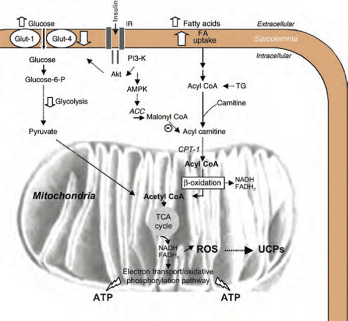
As an essential regulator of mitochondrial function and fatty acid metabolism, L-carnitine is relatively well known for its ability to improve muscle function in conditions such as cardiac ischemia, and is often used in the treatment of other muscular conditions such as fibromyalgia. The ability of carnitine to impact neurological function, however, is perhaps less well known. Surprisingly, there is a fairly well developed corpus around the use of acetyl-L-carnitine (ALC) in patients with neurological conditions ranging from age related dementia, substance withdrawal, depression, Down syndrome, attention deficient hyperactivity disorder, neuropathy, and encephalopathy secondary to liver failure (Arnold 2007, Janiri 2009, Malaguarnera 2008, Montgomery 2003, Pueschel 2006, Youle 2007, Zanardi 2006). This paper reviews the neuroprotective effects of ALC, with a narrowed focus on its effects in Alzheimer’s disease (AD), substance withdrawal, and toxic peripheral neuropathies.

Physiology

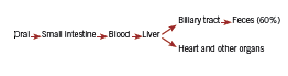
As reviewed previously, carnitine is a key factor in the transport of fatty acids between the cytoplasm and the mitochondria, facilitating production of ATP, and appears to be in especially high demand under ischemic conditions. Acetyl-L-carnitine passes the blood-brain barrier through an active transport process and is highly concentrated in the brain, especially in the hypothalamus (Thal 2000, Virmani 2004), and is therefore the main focus of carnitine research in the area of neurological disease.

ALC exerts its neuroprotective effect through a variety of mechanisms. According to Quatraro, ALC has analgesic effects by acutely increasing blood levels of the endogenous opioid peptide beta-endorphin, neurotropic effects by upregulating nerve growth factor receptors on the brain and preventing accumulation of lipofuscin, and metabolic effects by increasing the oxidative metabolism of neurons (Calvani 1992, Quatraro 1995). Pettegrew used magnetic resonance spectroscopy to show that administration of ALC normalized brain levels of high energy phosphates in patients with AD compared to healthy controls (1995). ALC also has antioxidant effects, increasing glutathione and decreasing malondialdehyde (Thal 2000). Finally, acetyl-L-carnitine is thought to have cholinergic effects through facilitation of intracellular acetylcholine synthesis; carnitine transports acetyl groups out of the mitochondra into the cytoplasm where they can be used for ACh production (Thal 2000, Virmani 2004).

Clinical Evidence: Cognitive Impairment and/ or Alzheieimer’s DiDisease

ALC has been shown to improve cognitive function in elderly patients without dementia. Two randomized double blind placebo controlled trials showed ALC to improve mental fatigue in patients over 70 and over 100 years of age, respectively, along with concomitant improvements in physical function (Malaguarnera 2008, 2007). A third trial reported significant improvements in cognitive function, memory, emotional affect, and mood when evaluated in 481 elderly patients without AD (Salvioli 1994).

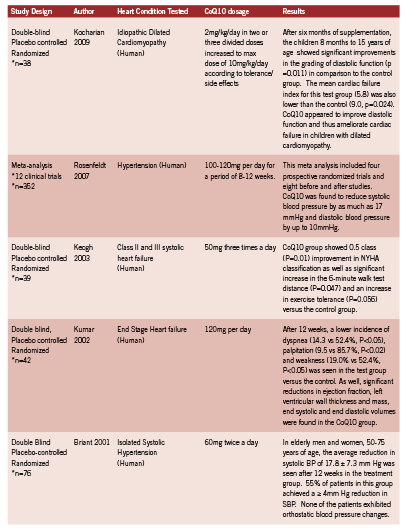
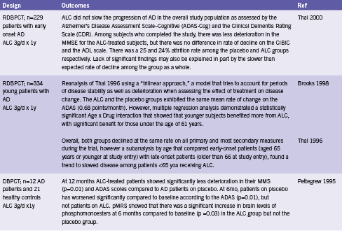
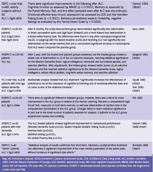
Two meta analyses have evaluated the effect of ALC for use in AD and/ or mild cognitive impairment (MCI) (Hudson 2003, Montgomery 2003). Montgomery included 21 randomized double blind placebo controlled trials assessing ALC for AD or MCI and concluded that ACL should be considered for the treatment of these conditions (2003). Doses ranged from 1.5 to 3.0 g/d for between 3 to 12 months. A combined effect size was calculated that incorporated both clinical and psychometric outcomes from the trials. There was significant benefit from treatment with ALC compared to placebo, effect size ES 0.201 (95% CI 0.107-0.295), and there was also a significant effect in the pooled Clinical Global Impression of Change, ES 0.32 (95% CI 0.18-0.47). ALC was well tolerated (Montgomery 2003).

The Cochrane review updated in 2008 included 16 randomized double blind placebo controlled trials (Hudson 2003). By contrast, this analysis found that while there was a statistically significant treatment effect in favour of ALC on the Clinical Global Impression at 12 and 24 wk (OR 1.90, 95% CI 1.31-2.76 and OR 2.33, 95% CI 1.31-4.14 respectively) this was not sustained at 52 weeks (OR 0.91, 95% CI 0.58-1.43). Similar effects were seen in pooled results for the Mini Mental State Exam (MMSE), leading authors to conclude that ALC may be of limited benefit.

This review excluded several studies that were included by Mongomery on the basis of poor reporting and/ or trial design. These trials were largely earlier trials out of Italy, so poor reporting standards may be to blame. In any case, the consequent discrepancy in included studies partially explains the divergent conclusions reached by the two studies. In addition, Mongomery utilized a unique composite effect measure that may not be directly comparable to those used by Hudson by pooling data across different assessment scales. However, it seems that the possibility of ALC benefiting at least a subset of patients with Alzheimer’s or MCI should not be ruled out. Hudson explains that the mechanisms by which ALC is absorbed and metabolized in the gut and liver “suggest that a large inter-individual variability should be expected in the general population. Studies have not taken this into account in selecting the dose of ALC, and this may be a source of error” (2003). Select trials of ALC for cognitive impairment are summarized in Table 1.

Table 1. Controlled Trials of Acetyl-L Carnitine for Alzheimer’s or Cognitive Impairment

Screen Shot 2014-07-01 at 9.38.19 AM

One uncontrolled trial found that the addition of ALC to the treatment of acetylcholinesterase inhibitor resistant patients with AD increased the response rate from 38% to 50% (p value not given) (Bianchetti 2003). ALC may therefore be useful for augmenting the effect of other therapies, even when it is not sufficient as a stand alone intervention.

Substance Withdrawal

A handful of trials have examined the potential effectiveness of ALC in ameliorating the symptoms of withdrawal from substances including methadone, cocaine, and alcohol. Janiri 2009 found that oral ALC 2g/d significantly decreased symptoms of methadone (opiate) withdrawal compared to placebo in 30 patients undergoing a three week detoxification program. Total symptom scores during the first five days were significantly lower in the ALC group (p<0.05), and in particular the following symptoms were improved: muscle tension and muscle spasm (p<0.05), insomnia (p<0.005), and feelings of coldness (p<0.05). Pain as assessed by the Huskisson analog scale was also “considerably lower” in the ALC group from after week one until the end of the study.

One study found no impact on symptoms of cocaine withdrawal or drug cravings (Reid 2005), while two RCTs reported significant improvements in cognitive impairment, mood, and anhedonia associated with chronic alcohol abuse in abstinent patients (Martinotti 2011, Tempesta 1990).

Peripheral Neuropathy 

ALC appears to protect nerves from the effects of toxic drugs such as antiretroviral therapy and chemotherapy, and from the damaging effects of diabetes. One RCT and three open trials have reported improvements in pain and/ or symptom scores from administration of ALC in patients with antiretroviral toxic neuropathy (Hart 2004, Osio 2006, Scarpini 1997, Youle 2007). Two open trials have found reductions in neuropathy grade and/ or symptomatic improvements in patients with neuropathy of chemotherapy (Bianchi 2005, Maestri 2005). Four RCTs and one open trial reported improvements in pain and/ or neuropathy grade when ALC was given to patients with diabetes (De Grandis 2002, Quatraro 1995, Sima 2005). These studies are summarized in Table 2.

Screen Shot 2014-07-01 at 9.38.33 AM
T able 2. Neuroprotective Properties of ALALC in Human Trials

Screen Shot 2014-07-01 at 9.38.43 AM

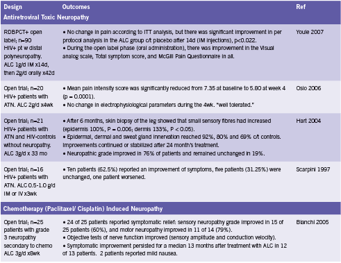
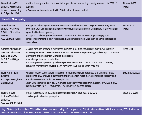
ALC has been shown to aid in the regeneration of damaged nerve tissue. Hart found that on biopsy, the skin of patients taking ALC for six months showed significant increases in the number of small sensory fibers, a 133% increase in the dermis, and 100% in the epidermis (2004). Furthermore, epidermal innervation reached up to 92% of that compared to healthy HIV- controls without neuropathy (Hart 2004). Sima demonstrated similar results in diabetic patients (2005). ALC has also been shown to improve of nerve conduction parameters, velocity and amplitude (Bianchi 2005, De Grandis 2002). Sima reported no effect on this outcome (2005), while Uzun found improvement in those patients with stage 1a diabetic neuropathy, but not those with stage 1b disease (2005).

Cerebrovascular Insufficiency

Finally, two small controlled trials have examined ALC in patients with cerebrovascular insufficiency (Arrigo 1990, Postiglione 1991). Intravenous administration of a single dose of ALC 1.5g increased cerebral blood flow compared to placebo (p<0.05) to the ischemic area but not the area corresponding to the stroke among 20 patients with a history of ischemic stroke when assessed by single proton emission computed tomography (SPECT) imaging (Postiglione 1991, Abstr). A cross over study in 12 patients undergoing rehab for cerebrovascular insufficiency reported that “significant differences between the drug [ALC] and placebo were found in memory, number and word tests and in the responses to simple stimuli and the performance of the maze test” (Arrigo 1990, Abstr).

Conclusion 

Acetyl-L-carnitine possesses analgesic, neuroprotective, metabolic, and cholinergic activities. Human studies have demonstrated clinical improvements associated with ALC use in a range of neurological conditions including Alzheimer’s disease and age related cognitive impairment, psychological and cognitive effects of chronic alcohol abuse, methodone withdrawal, toxic neuropathy of various etiologies, and chronic cerebrovascular insufficiency. Although there is some question as to the consistency of its effects between individual patients especially in AD, the majority of the evidence suggests that acetyl-L-carnitine would be a valuable intervention for use in the treatment of toxic or degenerative neurological conditions where permitted by jurisdiction of practice.

Acknowledgements 

The neuroprotective properties of ALC were first brought to this author’s attention by Paul Saunders ND, PhD.

References

Arnold LE, Amato A, Bozzolo H, Hollway J, Cook A, Ramadan Y, Crowl L, Zhang D, Thompson S, Testa G, Kliewer V, Wigal T, McBurnett K, Manos M. Acetyl-L-carnitine (ALC) in attention-deficit/hyperactivity disorder: a multi-site, placebo-controlled pilot trial. J Child Adolesc Psychopharmacol. 2007 Dec;17(6):791-802.

Arrigo A, Casale R, Buonocore M, Ciano C. Effects of acetyl-L-carnitine on reaction times in patients with cerebrovascular insufficiency. Int J Clin Pharmacol Res. 1990;10(1-2):133-7.

Bianchetti A, Rozzini R, Trabucchi M. Effects of acetyl-L-carnitine in Alzheimer’s disease patients unresponsive to acetylcholinesterase inhibitors. Curr Med Res Opin. 2003;19(4):350-3.

Bianchi G, Vitali G, Caraceni A, Ravaglia S, Capri G, Cundari S, Zanna C, Gianni L. Symptomatic and neurophysiological responses of paclitaxel- or cisplatin-induced neuropathy to oral acetyl-L-carnitine. Eur J Cancer. 2005 Aug;41(12):1746-50.

Bonavita E. Study of the efficacy and tolerability of L-acetylcarnitine therapy in the senile brain. Int J Clin Pharmacol Ther Toxicol. 1986 Sep;24(9):511-6.

Brooks JO 3rd, Yesavage JA, Carta A, Bravi D. Acetyl L-carnitine slows decline in younger patients with Alzheimer’s disease: a reanalysis of a double-blind, placebo-controlled study using the trilinear approach. Int Psychogeriatr. 1998 Jun;10(2):193-203.

Calvani M, Carta A, Caruso G, Benedetti N, Iannuccelli M. Action of acetyl-L-carnitine in neurodegeneration and Alzheimer’s disease. Ann N Y Acad Sci. 1992 Nov 21;663:483-6.

Cipolli C, Chiari G. [Effects of L-acetylcarnitine on mental deterioration in the aged: initial results]. Clin Ter. 1990 Mar 31;132(6 Suppl):479-510. [Abstr]

De Grandis D, Minardi C. Acetyl-L-carnitine (levacecarnine) in the treatment of diabetic neuropathy. A long-term, randomised, double-blind, placebo-controlled study. Drugs R D. 2002;3(4):223-31.

Hart AM, Wilson AD, Montovani C, Smith C, Johnson M, Terenghi G, Youle M. Acetyl-l-carnitine: a pathogenesis based treatment for HIV-associated antiretroviral toxic neuropathy. AIDS. 2004 Jul 23;18(11):1549-60.

Hudson S, Tabet N. Acetyl-L-carnitine for dementia. Cochrane Database Syst Rev. 2003;(2):CD003158. Review. PubMed PMID: 12804452.

Janiri L, Martinotti G, Tonioni F, Ghelardini C, Nicolai R, Galeotti N, Mosconi L, Calvani M, Bartolini A, Iannoni E. Acetyl-L-carnitine in the management of pain during methadone withdrawal syndrome. Clin Neuropharmacol. 2009 Jan-Feb;32(1):35-40.

Maestri A, De Pasquale Ceratti A, Cundari S, Zanna C, Cortesi E, Crinò L. A pilot study on the effect of acetyl-L-carnitine in paclitaxel- and cisplatin-induced peripheral neuropathy. Tumori. 2005 Mar-Apr;91(2):135-8.

Malaguarnera M, Cammalleri L, Gargante MP, Vacante M, Colonna V, Motta M. L-Carnitine treatment reduces severity of physical and mental fatigue and increases cognitive functions in centenarians: a randomized and controlled clinical trial. Am J Clin Nutr. 2007 Dec;86(6):1738-44.

Malaguarnera M, Gargante MP, Cristaldi E, Vacante M, Risino C, Cammalleri L, Pennisi G, Rampello L. Acetyl-L-carnitine treatment in minimal hepatic encephalopathy. Dig Dis Sci. 2008 Nov;53(11):3018-25.

Malaguarnera M, Gargante MP, Cristaldi E, Colonna V, Messano M, Koverech A, Neri S, Vacante M, Cammalleri L, Motta M. Acetyl L-carnitine (ALC) treatment in elderly patients with fatigue. Arch Gerontol Geriatr. 2008 Mar-Apr;46(2):181-90.

Martinotti G, Andreoli S, Reina D, Di Nicola M, Ortolani I, Tedeschi D, Fanella F, Pozzi G, Iannoni E, D’Iddio S, Prof LJ. Acetyl-l-Carnitine in the treatment of anhedonia, melancholic and negative symptoms in alcohol dependent subjects. Prog Neuropsychopharmacol Biol Psychiatry. 2011 Jan 20. [Epub ahead of print]

Montgomery SA, Thal LJ, Amrein R. Meta-analysis of double blind randomized controlled clinical trials of acetyl-L-carnitine versus placebo in the treatment of mild cognitive impairment and mild Alzheimer’s disease. Int Clin Psychopharmacol. 2003 Mar;18(2):61-71.

Neri M, Cortelloni C, De Vreese L. [Methodology of a clinical controlled study of L-acetylcarnitine]. Clin Ter. 1990 Mar 31;132(6 Suppl):457-68. Italian.

Osio M, Muscia F, Zampini L, Nascimbene C, Mailland E, Cargnel A, Mariani C. Acetyl-l-carnitine in the treatment of painful antiretroviral toxic neuropathy in human immunodeficiency virus patients: an open label study. J Peripher Nerv Syst. 2006 Mar;11(1):72-6.

Passeri M, Cucinotta D, Bonati PA, Iannuccelli M, Parnetti L, Senin U. Acetyl-L-carnitine in the treatment of mildly demented elderly patients. Int J Clin Pharmacol Res. 1990;10(1-2):75-9.

Passeri M, Iannuccelli M, Ciotti G, Bonati PA, Nolfe G, Cucinotta D. Mental impairment in aging: selection of patients, methods of evaluation and therapeutic possibilities of acetyl-L-carnitine. Int J Clin Pharmacol Res. 1988;8(5):367-76.

Pettegrew JW, Klunk WE, Panchalingam K, Kanfer JN, McClure RJ. Clinical and neurochemical effects of acetyl-L-carnitine in Alzheimer’s disease. Neurobiol Aging. 1995 Jan-Feb;16(1):1-4.

Postiglione A, Soricelli A, Cicerano U, Mansi L, De Chiara S, Gallotta G, Schettini G, Salvatore M. Effect of acute administration of L-acetyl carnitine on cerebral blood flow in patients with chronic cerebral infarct. Pharmacol Res. 1991 Apr;23(3):241-6.

Pueschel SM. The effect of acetyl-L-carnitine administration on persons with Down syndrome. Res Dev Disabil. 2006 Nov-Dec;27(6):599-604.

Quatraro A, Roca P, Donzella C, Acampora R, Marfella R, Giugliano D. Acetyl-L-carnitine for symptomatic diabetic neuropathy. Diabetologia. 1995 Jan;38(1):123.

Rai G, Wright G, Scott L, Beston B, Rest J, Exton-Smith AN. Double-blind, placebo controlled study of acetyl-l-carnitine in patients with Alzheimer’s dementia. Curr Med Res Opin. 1990;11(10):638-47.

Reid MS, Casadonte P, Baker S, Sanfilipo M, Braunstein D, Hitzemann R, Montgomery A, Majewska D, Robinson J, Rotrosen J. A placebo-controlled screening trial of olanzapine, valproate, and coenzyme Q10/L-carnitine for the treatment of cocaine dependence. Addiction. 2005 Mar;100 Suppl 1:43-57.

Salvioli G, Neri M. L-acetylcarnitine treatment of mental decline in the elderly. Drugs Exp Clin Res. 1994;20(4):169-76.

Sano M, Bell K, Cote L, Dooneief G, Lawton A, Legler L, Marder K, Naini A, Stern Y, Mayeux R. Double-blind parallel design pilot study of acetyl levocarnitine in patients with Alzheimer’s disease. Arch Neurol. 1992 Nov;49(11):1137-41.

Scarpini E, Sacilotto G, Baron P, Cusini M, Scarlato G. Effect of acetyl-L-carnitine in the treatment of painful peripheral neuropathies in HIV+ patients. J Peripher Nerv Syst. 1997;2(3):250-2.

Sima AA, Calvani M, Mehra M, Amato A; Acetyl-L-Carnitine Study Group. Acetyl-L-carnitine improves pain, nerve regeneration, and vibratory perception in patients with chronic diabetic neuropathy: an analysis of two randomized placebo-controlled trials. Diabetes Care. 2005 Jan;28(1):89-94.

Spagnoli A, Lucca U, Menasce G, Bandera L, Cizza G, Forloni G, Tettamanti M, Frattura L, Tiraboschi P, Comelli M, et al. Long-term acetyl-L-carnitine treatment in Alzheimer’s disease. Neurology. 1991 Nov;41(11):1726-32.

Tempesta E, Troncon R, Janiri L, Colusso L, Riscica P, Saraceni G, Gesmundo E, Calvani M, Benedetti N, Pola P. Role of acetyl-L-carnitine in the treatment of cognitive deficit in chronic alcoholism. Int J Clin Pharmacol Res. 1990;10(1-2):101-7.

Thal LJ, Calvani M, Amato A, Carta A. A 1-year controlled trial of acetyl-l-carnitine in early-onset AD. Neurology. 2000 Sep 26;55(6):805-10.

Thal LJ, Carta A, Clarke WR, Ferris SH, Friedland RP, Petersen RC, Pettegrew JW, Pfeiffer E, Raskind MA, Sano M, Tuszynski MH, Woolson RF. A 1-year multicenter placebo-controlled study of acetyl-L-carnitine in patients with Alzheimer’s disease. Neurology. 1996 Sep;47(3):705-11.

Uzun N, Sarikaya S, Uluduz D, Aydin A. Peripheric and automatic neuropathy in children with type 1 diabetes mellitus: the effect of L-carnitine treatment on the peripheral and autonomic nervous system. Electromyogr Clin Neurophysiol. 2005 Sep-Oct;45(6):343-51.

Virmani A, Binienda Z. Role of carnitine esters in brain neuropathology. Mol Aspects Med. 2004 Oct-Dec;25(5-6):533-49.

Youle M, Osio M; ALCAR Study Group. A double-blind, parallel-group, placebo-controlled, multicentre study of acetyl L-carnitine in the symptomatic treatment of antiretroviral toxic neuropathy in patients with HIV-1 infection. HIV Med. 2007 May;8(4):241-50.

Zanardi R, Smeraldi E. A double-blind, randomised, controlled clinical trial of acetyl-L-carnitine vs. amisulpride in the treatment of dysthymia. Eur Neuropsychopharmacol. 2006 May;16(4):281-7.

CoEnzyme Q10

0

CoEnzyme Q10

Cardiovascular applications

Introduction

In 2007, heart disease was one of the two leading causes of death for Canadians and was responsible for 22% of the 235,217 deaths (Statistics Canada 2010). Heart disease includes heart failure, angina, cardiomyopathy, coronary artery disease, valve disorders and other conditions related to the heart. ese staggering numbers call for the best possible treatments for heart disease whereupon Coenzyme Q10 (CoQ10) has appeared as a promising therapy.

Heart Facts e adult human heart weighs from 200 to 425g and is able to convert chemical energy to mechanical energy extremely effi ciently. To put it in numbers, it is capable of pumping out fi ve liters of blood every minute, 7200 liters per day and over 2.6 million liters every year. In order to execute this, the heart hydrolyzes an estimated six kilograms of adenosine-5’-triphosphate (ATP) per day (Soukoulis 2009).

High blood pressure (BP) is one of the most important cardiovascular risk factors worldwide. Approximately two-thirds of patients do not achieve optimal BP control using drug therapy (Yusuf 2004). Other interventions are extremely important, as a reduction of 5mmHg in systolic BP has been associated with a 7% reduction in all-cause mortality (Whelton 2002).

The consequences of heart failure are concerning and despite sophisticated diagnostic techniques and treatments, the risk of death within fi ve years of diagnosis is greater than 50% (Dunn 2009). CoQ10 levels in the blood have been found to be an independent predictor of mortality in congestive heart failure (Molyneux 2008).

About CoQ10

First isolated in 1957 from beef mitochondria, CoQ10, or ubiquinone is highly concentrated in heart-muscle cells due to their increased energy requirements (Crane 1989). In addition to its role in the formation of ATP, CoQ10 serves to delay or prevent lipid peroxidation and enhances cell membrane stabilization (Sarter 2002, Shah 2007). Due to the fact that mitochondria are particularly vulnerable to oxidative damage, mitochondria targeted antioxidants like CoQ10 can be an effective therapeutic strategy in preventing or reducing the progression of cardiovascular and other disorders (Graham 2009).

CoQ10 is mostly found in active organs like the heart where a substantial decline can be observed with increasing age (Pravst 2010). An adult human body has approximately 2 grams of CoQ10 and 0.5 grams must be replaced daily making the average turnover rate in the body around 4 days (Pravst 2010). Human intervention trials have found supplemental CoQ10 to be effective in cardiovascular disorders like cardiomyopathy, hypertension, angina pectoris, atherosclerosis, ischemic heart disease, cardiovascular surgery, hypertension, valvular heart diseases and myocardial infarctions (Hadj 2007, Kumar 2009).

Sources

Common sources for CoQ10 are beef, poultry, broccoli, soya oil, fish oils, peanuts, sardines, and mackerel. However, an average dietary amount of CoQ10 is only 2-6mg/day, which is inadequate to provide levels in the body required to be beneficial in pathological states (Kumar 2009, Pravst 2010).

Screen Shot 2014-07-07 at 2.36.18 PM

Metabolism and Excretion

CoQ10 is absorbed from the small intestine and transferred into the blood circulation. It is then carried to the liver where it undergoes biotransformation and is primarily excreted through the bile duct. In this process, only a fraction of the ingested amount is carried to other organs like the heart, adrenal glands, kidneys and lungs (Ozaki 2010, Wyman 2010). Approximately 60% of an oral dose is eliminated in the feces during chronic oral administration and the elimination half-life is about 34 hours (Greenberg 1990). Due to its excretion pathway, patients with biliary obstruction or hepatic impairment may accumulate CoQ10 in their bodies (Ozaki 2010, Wyman 2010).

Absorbability and Dosage

Due to its lipophilic nature, CoQ10 is best absorbed with meals or in emulsified form. Divided dosing also maximizes absorbability while minimizing potential side effects (Ozaki 2010).

What form of CoQ10 is best? According to Hosoe (2007), ubiquinol, the reduced form of CoQ10, is two times more absorbable than ubiquinone. In support of ubiquinol, Langsjoen (2008) found that ubiquinol dramatically improved absorption and was correlated to improvements in CoQ10 levels and positive clinical outcomes that were not possible with the use of ubiquinone at even up to 900mg/ day. However, it has also been reported that the form of CoQ10 ingested may not be so important. According to Pravst (2010), ubiquinone appears to be reduced during or following absorption in the intestine, and as a result more than 95% of CoQ10 in circulation exists in the more active ubiquinol form after its ingestion. Thus, the answer to this question is still in contention.

The recommended daily dose of CoQ10 ranges from 30 to 100mg per day for healthy people (Pravst 2010) or 60 to 200mg per day in treating different conditions (Kumar, 2009). A maximum dosage of 1200mg has recently been suggested for adult intake as it has been found to be well tolerated and safe (Hathcock 2006).

Normal blood levels range from 0.7 to 1.0 ug/ml; doses of 30-60mg can be used to prevent CoQ10 deficiency and to maintain normal serum concentrations. However, clinically useful levels necessitate above normal CoQ10 blood levels that may be two to four times higher. In fact, 450mg of CoQ10 a day was found to achieve a plasma level of 4ug/ml and was much more successful in reversing the course of severe heart failure (Kumar 2009).

These increased levels may take days or months to achieve. Oral administration of 100mg/day of CoQ10 for two to eight months resulted in an increase of 20-85% in myocardial CoQ10 levels in patients with cardiomyopathy (Folkers 1985, Sarter 2002). Due to this slow plasma increase of CoQ10, clinical improvement is normally seen one to four weeks after initiation of treatment and it may take months to reach maximal clinical benefit (Kumar 2009).

Mechanism of action

CoQ10 appears to work in several ways including targeting expression of multiple genes, such as those involved in intermediary metabolism and cell signaling. This gene regulation and control of metabolism may explain many of the cardiovascular and other actions of CoQ10 (Rosenfeldt 2007). The beneficial effect of CoQ10 in hypertensive cases is a result of decreased peripheral resistance that is due to its direct action on the vascular wall. Free radicals inactivate nitric oxide (NO), thus preventing NOmediated relaxation of the smooth muscle layer of the vascular wall. By acting as a free radical scavenger, CoQ10 is able to prevent vasoconstriction and the resulting increase in blood pressure (Ankola 2007). With regards to coronary heart failure and myocardial infarction, CoQ10 is hypothesized to benefit these states through its direct impact on energy production by mitochondria, improving ATP availability for the failing heart (Kumar 2009, Crane 2001).

Untitled-27

Side Effects

A long-term noncomparative study was conducted in 173 Italian centers to specifically look at safety and efficacy of CoQ10 in 2664 patients with congestive heart failure (Baggio 1994). In this study, preexisting adverse reactions were taken at baseline and then recorded again after three months of treatment with only 36 patients (1.3%) complaining of side effects mainly to do with mild gastrointestinal upset (Baggio 1994). The maximum dose in this study was 150mg per day. However, CoQ10 supplementation has been found to be free of side effects at dosages up to 600mg/day and well tolerated and safe up to 1200mg/day (Hathcock 2006). Aside from possible abdominal discomfort, nausea, vomiting, diarrhea and anorexia, allergic rash and headache have also been reported in rare cases. It is reported that CoQ10’s antiplatelet effect may increase the risk of bleeding, especially in those on antiplatelet medication (Wyman 2010). However, due to its vitamin K like action it may also act to oppose the anticoagulant effects of warfarin (Jelin 2009).

Clinical evidence

Research for CoQ10 has resulted in many positive conclusions that include Langsjoen’s study published in 1994 illustrating the usefulness of CoQ10 in clinical cardiology. It tested the use of CoQ10 in 424 patients with different types of myocardial diseases. Patients were treated with an average of 240mg of CoQ10 per day and followed an average of approximately a year and a half. Significant improvement in New York Heart Association functional classification (Table 1) was seen. 58% of patients improved by one NYHA class, 28% improved by two classes, 1.2% by three NYHA classes. Within a month, myocardial function became measurably improved. By six months, maximal improvement was usually obtained and this improvement was sustained in the majority of patients. 43% stopped between one and three drugs and only 6% required the addition of one drug. The withdrawal of CoQ10 supplementation resulted in a measurable decline in myocardial function within 1 month and a regression to pretreatment measurements within three to six months (Langsjoen 1994a, Langsjoen 1994b).

Table 1 New York Heart Association (NYHA) Classification of Functional Capacity (Chavey 2001)
Table 1
New York Heart Association (NYHA)
Classification of Functional Capacity
(Chavey 2001)

In 1994, Baggio published the largest open trial in heart failure, which included 2664 patients treated with up to 150 mg of CoQ10 per day. At the end of three-months, improvements were seen in vertigo (73.1%), subjective arrhythmia (63.4%), insomnia (62.8%), cyanosis (78.1%), edema (78.6%), pulmonary rales (77.8%), hepatomegaly (49.3%), jugular reflux (71. 8%), dyspnea (52. 7%), palpitations (75.4%), sweating (79.8%), and nocturia (53.6%). Over half (54%) of patients had improvement of at least three symptoms. Furthermore, 89.7% entered as NYHA class II moved up to a NYHA class I classification and 28.8% of patients entered as NYHA class III classification moved up to a class II classification (Baggio 1994).

The elderly population is of particular interest as using antihypertensive agents can be problematic because of the increased incidence of postural hypotension with resulting risk of falls and associated morbidity. Two large clinical trials in 1991 and 1997 respectively confirmed that in adults over the age of 60 with isolated systolic hypertension (systolic blood pressure >140, diastolic blood pressure <90), reducing systolic blood pressure by 20 mm Hg reduces the incidence of stroke, heart failure and mortality (SHEP Cooperative Research Group 1991, Rosenfeld 1997). Clinical trials of CoQ10 in hypertension, as well as trials of CoQ10 in cardiovascular pathologies, are summarized in Table 2.

Table 2 Important clinical trials of CoQ10 in hypertension and cardiovascular pathologies

Clinical implications The use of CoQ10 in patients with heart disease may not only be useful but imperative in achieving clinically relevant improvements in heart disease patients. While more research in this area is needed with larger “n” numbers in order to statistically better quantify its merits, much research thus far points to positive outcomes in its utilization. However, its low side effect profile and non-toxic nature makes CoQ10 a viable treatment for all age groups and across a broad range of cardiovascular pathologies.

Following positive results outlined here and elsewhere, countries like Japan, Hungary, Italy, Norway and Denmark now grant licensed prescription of CoQ10 for heart failure and ischemic heart disease (Pepe 2007), a move that shows its valued acceptance and one that encourages a more prominent and wide-spread use of CoQ10.

Untitled-28

References

Ankola DD, Viswanad B, Bhardwaj V, Ramarao P, Kumar MN. Development of potent oral nanoparticulate formulation of coenzyme Q10 for treatment of hypertension: can the simple nutritional supplements be used as first line therapeutic agents for prophylaxis/therapy? Eur J Pharm Biopharm. 2007;67(2):361-9. Epub 2007.

Baggio E, Gandini R, Plancher AC, Passeri M, Carmosino G. Italian multicenter study on the safety and efficacy of coenzyme Q10 as adjunctive therapy in heart failure. CoQ10 Drug Surveillance Investigators. Mol Aspects Med 1994;15(Suppl):S287-94.

Burke BE, Neuenschwander R, Olson RD. Randomized, double-blind, placebo-controlled trial of coenzyme Q10 in isolated systolic hypertension. South Med J. 2001;94(11):1112-7.

Chavey WE 2nd, Blaum CS, Bleske BE, Harrison RV, Kesterson S, Nicklas JM. Guideline for the management of heart failure caused by systolic dysfunction: Part I. Guideline development, etiology and diagnosis. Am Fam Physician. 2001;64(5):769-74.

Crane, FL, Hatefi Y, Lester RI, Widmer C. Isolation of a quinone from beef heart mitochondria. 1957. Biochim Biophys Acta. 1989;1000:362-3.

Crane FL. Biochemical functions of coenzyme Q10. J Am Coll Nutr. 2001;20(6):591-8.

Dunn SP, Bleske B, Dorsch M, Macaulay T, Van Tassell B, Vardeny O. Nutrition and heart failure: impact of drug therapies and management strategies. Nutr Clin Pract. 2009;24(1):60- 75.

Folkers K, Vadhanavikit S, Mortensen SA. Biochemical rationale and myocardial tissue data on the effective therapy of cardiomyopathy with coenzyme Q10. Proc Natl Acad Sci 1985;82:901–904.

Graham D, Huynh NN, Hamilton CA, Beattie E, Smith RA, Cochemé HM, Murphy MP, Dominiczak AF. Mitochondria-targeted antioxidant MitoQ10 improves endothelial function and attenuates cardiac hypertrophy. Hypertension. 2009;54(2):322-8. Epub 2009.

Greenberg S, Frishman WH. Co-enzyme Q10: a new drug for cardiovascular disease. J Clin Pharmacol 1990;30:596–608.

Hadj A, Pepe S, Rosenfeldt F. The clinical application of metabolic therapy for cardiovascular disease. Heart Lung Circ. 2007;16 Suppl 3:S56-64. Epub 2007.

Hathcock, J. N., & Shao, A. (2006). Risk assessment for coenzyme Q10 (ubiquinone). Regul Toxicol Pharmacol 45, 282–288.

Hosoe K, Kitano M, Kishida H, Kubo H, Fujii K, Kitahara M. Study on safety and bioavailability of ubiquinol after single and 4-week multiple oral administration to healthy volunteers. Regul Toxicol Pharmacol 2007;7(1):19–28.

Jelin JM, Gregory PJ, et al. Natural medicines comprehensive database/compiled by the editors of Pharmacist’s Letter, Prescriber’s Letter. 11th ed. Stockton, CA: Therapeutic Research Faculty; 2009:452–457.

Keogh A, Fenton S, Leslie C, Aboyoun C, Macdonald P, Zhao YC, Bailey M, Rosenfeldt F. Randomised double-blind, placebo-controlled trial of coenzyme Q , therapy in class II and III systolic heart failure. Heart Lung Circ. 2003;12(3):135-41.

Kocharian A, Shabanian R, Rafiei-Khorgami M, Kiani A, Heidari-Bateni G. Coenzyme Q10 improves diastolic function in children with idiopathic dilated cardiomyopathy. Cardiol Young. 2009;19(5):501-6. Epub 2009.

Kumar A, Kartikey NS, Niaz MA, Singh RB. Effect of Q gel (coenzyme Q10) in patients with end stage heart failure targetted for heart transplantation. 2002. Third Conference of the International Coenzyme Q10 Association, London, UK.

Kumar A, Kaur H, Devi P, Mohan V. Role of coenzyme Q10 (CoQ10) in cardiac disease, hypertension and Meniere-like syndrome. Pharmacol Ther. 2009;124(3):259-68. Epub 2009.

Langsjoen H, Langsjoen P, Langsjoen P, Willis R, Folkers K. Usefulness of coenzyme Q10 in clinical cardiology: a long-term study. Mol Aspects Med 1994a;15 Suppl:s165–s175.

Langsjoen P, Willisb R, Folkers K. Treatment of essential hypertension with coenzyme Q10. Mol Aspects Med 1994b;15:262–272.

Langsjoen PH, Langsjoen AM. Biofactors. Supplemental ubiquinol in patients with advanced congestive heart failure. 2008;32(1-4):119-28.

Molyneux SL, Young JM, Florkowski CM, Lever M, George PM. Coenzyme Q10: is there a clinical role and a case for measurement? Clin Biochem Rev 2008;29:71–82.

Ozaki A, Muromachi A, Sumi M, Sakai Y, Morishita K, Okamoto T. Emulsification of coenzyme Q10 using gum arabic increases bioavailability in rats and human and improves foodprocessing suitability. J Nutr Sci Vitaminol (Tokyo). 2010;56(1):41-7.

Pepe S, Marasco SF, Haas SJ, Sheeran FL, Krum H, Rosenfeldt FL. Coenzyme Q10 in cardiovascular disease. Mitochondrion. 2007;7 Suppl:S154-67. Epub 2007.

Pravst I, Zmitek K, Zmitek J. Coenzyme Q10 contents in foods and fortification strategies. Crit Rev Food Sci Nutr. 2010;50(4):269-80.

Rosenfeld J, Rodicio JL, Tuomilehto J, Zanchetti A. Randomized double-blind comparison of placebo and active treatment for older patients with isolated systolic hypertension. The Systolic Hypertension in Europe (Syst-Eur) Trial Investigators. Lancet 1997;350:757-764.

Rosenfeldt FL, Haas SJ, Krum H, Hadj A, Ng K, Leong JY, Watts GF. Coenzyme Q10 in the treatment of hypertension: a meta-analysis of the clinical trials. J Hum Hypertens. 2007;21(4):297-306. Epub 2007.

Sarter B. Coenzyme Q10 and cardiovascular disease: a review. J Cardiovasc Nurs. 2002;16(4):9-20.

Shah SA, Sander S, Cios D, Lipeika J, Kluger J, White CM. Electrocardiographic and hemodynamic effects of coenzyme Q10 in healthy individuals: a double-blind, randomized controlled trial. Ann Pharmacother. 2007;41(3):420-5. Epub 2007.

SHEP Cooperative Research Group. Prevention of stroke by antihypertensive drug treatment in older persons with isolated systolic hypertention. Final results of the Systolic Hypertension in the Elderly Program (SHEP). JAMA 1991;265:3255-3264.

Staessen JA, Fagard R, Thijs L, Celis H, Arabidze GG, Birkenhäger WH, Bulpitt CJ, de Leeuw PW, Dollery CT, Fletcher AE, Forette F, Leonetti G, Nachev C, O’Brien ET,

Soukoulis V, Dihu JB, Sole M, Anker SD, Cleland J, Fonarow GC, Metra M, Pasini E, Strzelczyk T, Taegtmeyer H, Gheorghiade M. Micronutrient deficiencies an unmet need in heart failure. J Am Coll Cardiol. 2009;54(18):1660-73.

Statistics Canada. Leading causes of death. http://www.statcan.gc.ca/daily-quotidien/101130/ dq101130b-eng.htm. Accessed December 2010.

Whelton PK, He J, Appel LJ, Cutler JA, Havas S, Kotchen TA, et al. Primary prevention of hypertension: clinical and public health advisory from the National High Blood Pressure Education Program. JAMA 2002;288(15):1882-8.

Wyman M, Leonard M, Morledge T. Coenzyme Q10: a therapy for hypertension and statininduced myalgia? Cleve Clin J Med. 2010;77(7):435-42.

Yusuf S, Hawken S, Ounpuu S, Dans T, Avezum A, Lanas F. Effect of potentially modifiable risk factors associated with myocardial infarction in 52 countries (the INTERHEART study): case-control study. Lancet 2004;364(9438):937-52.

L- Carnitine

0

L- Carnitine

Heidi Fritz MA, ND

Introduction

L-carnitine is a trimethylated amino acid- derivative whose most well described biological function is as a cofactor of fatty acid metabolism (Alt Med Rev 2005). L-carnitine is a key component of the intracellular shuttle system that transports free fatty acids from the cytosol and across the mitochondrial membrane where they undergo beta oxidation and transformation to ATP (Karlic 2004). Long chain fatty acids represent up to 80% of the body’s energy sources during most physiological states with the exception of high intensity exercise (Karlic 2004) and are the preferred substrate in cardiac muscle (Carvajal 2003). L-carnitine also impacts other cellular energy- producing pathways (Mate 2010). Optimization of mitochondrial metabolism through supplementation with L-carnitine, which is known to concentrate in cardiac and skeletal muscle (Flanagan 2010), could be an effective intervention for a variety of conditions characterized by fatigue, pain, and impaired muscle function. This paper focuses on the evidence base for L-carnitine in cardiovascular medicine.

Physiology

L-carnitine is considered to be conditionally essential, meaning that although there are rare genetic conditions causing primary deficiency, and secondary deficiency resulting commonly from hemodialysis in patients with kidney disease, most healthy individuals can synthesize L-carnitine from dietary lysine and methionine and cofactors vitamin C, iron, pyroxidine, and niacin (Flanagan 2010, Mate 2010). Despite this, alterations of L-carnitine metabolism are common in cardiovascular disease, most likely due to a combination of increased utilization and the inability of myocardiocytes to synthesize carnitine endogenously.

Cardiac ischemia is well recognized in the literature to be accompanied by a rapid depletion in myocardial carnitine content and a concurrent rise in intracellular long chain free fatty acids (Gurlek 2000, Regitz 1990A, Regitz 1990B, Tarantini 2006). In patients with heart failure, serum carnitine has been correlated with impaired left ventricular systolic function as measured by echocardiography (El-Aroussy 2000). During ischemia L-carnitine is thought to offset rising concentrations of free fatty acids by facilitating mitochondrial uptake and utilization, thereby preventing the damaging effects of elevated free fatty acids: membrane damage with consequent cell swelling and microvascular compression, arrhythmias, and metabolic inefficiency with consequent deterioration of myocardial function (Tarantini 2006).

In addition to its facilitating fatty acid transport into the mitochondria, L-carnitine performs a second key metabolic function: the removal from the mitochondria of short and medium chain fatty acids (acetyl groups) formed as products of beta oxidation and bound to CoA as acetyl-CoA (Mate 2010). Mitochondrial accumulation of this byproduct is toxic, inhibiting pyruvate dehydrogenase activity and glucose oxidation. On the other hand, the removal of acetyl groups and consequent increase in free CoA stimulates energy production through the Kreb’s cycle (Mate 2010). In this way, L-carnitine can regulate both fatty acid and glucose metabolism in ischemic cardiac tissue. See Figure 1. Importantly, since myocardium is unable to synthesize carnitine itself, exogenous administration may be necessary to restore optimal levels and metabolic function.

Untitled-25

Human Evidence

Oral and intravenous L-carnitine has been shown to be benefi cial for a number of cardiovascular conditions including angina (Bartels 1996, Cacciatore 1991), acute myocardial infarction (Gurlek 2000), chronic heart failure (Rizos 2000), and peripheral vascular disease (Brevetti 1999); because it is more accessible, this summary focuses on oral L-carnitine in particular. e typical dosage is 2g per day in divided doses (Gurlek 2000, Singh 1996); some studies have used up to 6g per day (Iliceto 1995). No adverse events beyond mild gastrointestinal upset, which was rare, were reported (Kumar 2007). It should be noted that most of the studies cited here included patients also receiving standard medications for their conditions, and that L-carnitine was studied as an “add on” therapy; space constraints limit the detail in which this can be described for each study.

Angina ere are at least eight controlled human trials of oral L-carnitine for the treatment of angina. Of these, all found benefi t (Bartels 1995, Bartels 1996, Cacciatore 1991, Cherchi 1985, Cherchi 1990, Iyer 2000, Kamikawa 1984, Lagioia 1992, McMackin 2007). L carnitine has been shown to:

• Improve cardiac function (eg. cardiac output, maximal heart rate) during exercise and improve overall exercise performance (eg. exercise time, exercise workload) despite having no eff ect on myocardial oxygen requirements (Bartels 1996, Bartels 1995, Cacciatore 1991, Cherchi 1985, Iyer 2000, Kamikawa 1984, Lagioia 1992);

• Reduce myocardial ischemia upon exercise testing, measured as the time to ST-segment depression or the degree of ST-depression on ECG (Bartels 1996, Bartels 1995, Cacciatore 1991, Cherchi 1985, Cherchi 1990, Kamikawa 1984, Lagioia 1992);

• Reverse angina, with a number of patients becoming angina- free during treatment (up to 22.7% of patients compared to 9.1% in the placebo group according to Cherchi 1985, 2 out of 12 patients according to Kamikawa 1984); • Decrease frequency of angina attacks by up to 70% and nitroglycerin consumption by 57% according to Bartels 1996, comparable to treatment with diltiazem;

• One additional study in patients without angina but with established coronary artery disease showed L-carnitine to increase brachial artery diameter by 2.3%, consistent with reduced arterial tone (McMackin 2007).

L-carnitine exerts these eff ects without reducing myocardial oxygen demand, as do select cardiac medications such as beta blockers. Bartels showed that: “[proprionyl-L-carnitine] prevents ischemia-induced ventricular dysfunction, not by aff ecting the myocardial oxygen supply-demand ratio but as a result of its intrinsic metabolic actions, increasing pyruvate dehydrogenase activity and fl ux through the citric acid cycle” (1994).

Acute Myocardial Infarction

Eight controlled trials have examined the eff ect of L-carnitine in the treatment of acute myocardial infarction (AMI), often with intravenous administration for an initial period, followed by long term oral administration. ese are summarized in Table 1. When added to standard therapies, L-carnitine has been found to:

• Improve myocardial pumping ability: increasing left ventricular ejection fraction (LVEF) (Gurlek 2000), and end- diastolic and end- systolic volumes (Iliceto 1995)

• Reduce myocardial damage: reduced infarct size when administered acutely (Singh 1996), and reduced deleterious left ventricular remodeling and dilation when administered acutely and long term (Iliceto 1995)

• Decrease angina following MI by up to half that experienced by patients receiving placebo (Davini 1992, Singh 1996)

• Decrease incidence of arrhythmias following MI (Mondillo 1995, Singh 1996)

• Reduce number of second cardiac events to 15.6%, compared to 26% in placebo (Singh 1996)

• Reduce overall mortality, with 1.2% rate reported in LC group compared to 12.5% in the placebo group according to Davini 1992. Reduced mortality also demonstrated by De Pasquale (1995).

Two large randomized controlled trials have been conducted by Italian researchers. The CEDIM1 (Carnitine Ecocardiografia Digitalizzata Infarto Miocardico) and CEDIM2 trials investigated the use of intravenous L-carnitine 9g/d for the first five days after AMI, followed by 4 to 6 g/d orally for 6 to 12 months in 472 and 2230 patients respectively (Iliceto 1995, Tarantini 2006). Although the studies differed in their long term (6-12 months) findings, both demonstrate that L-carnitine can reduce early mortality post AMI. CEDIM2 found significantly reduced early mortality, risk of death at 5 days HR 0.61 (95% CI 0.37-0.98), and upon reanalysis, it was found that much of the apparent benefit seen in CEDIM1 was also due to lower early mortality (Tarantini 2006).

Of the eight studies, seven found significant positive effects (Davini 1992, De Pasquale 1995, Gurlek 2000, Iliceto 1995, Mondillo 1995, Singh 1996, Tarantini 2006). Iyer found no effect on cardiac function in a smaller trial of 60 patients using L-carnitine 6g/d intravenously for the first 7 days, then 3g/d orally for 3 months, and it has been suggested that this may be due to the smaller trial size compared to CEDIM 1 and 2, or the lower dosage used (1999).

Heart Failure

Nine controlled trials have investigated L-carnitine for patients with chronic/ congestive heart failure (CHF) (Anand 1998, Caponetto 1994, Ghidini 1988, Kumar 2007, Loster 1999, Mancini 1992, No authors 1999, Pucciarelli 1992, Rizos 2000). L-carnitine has been shown to:

• Improve exercise performance (maximum exercise time) (Caponetto 1994, Kumar 2007, Loster 1999, Mancini 1992, No authors 1999, Pucciarelli 1992, Rizos 2000)

• Improve cardiac function during exercise (eg. peak exercise heart rate, ejection fraction) (Anand 1998, Mancini 1992, No authors 1999)

• Increase cardiac pumping ability (eg. cardiac output, stroke volume index, left ventricular ejection fraction, pulmonary blood pressure) (Caponetto 1994, Pucciarelli 1992, Rizos 2000)

• Decrease left ventricular size (Anand 1998)

• Decrease signs and symptoms associated with CHF, including dyspnea, palpitations, fatigue, edema, and improved diuresis (Ghidini 1988, Kumar 2007)

• Reduce long term mortality: in one study 3-year mortality was 3% for patients receiving L-carnitine, compared to 18% for those on placebo (p<0.04) (Rizos 2000).

Interestingly, Loster found that the beneficial effects of L-carnitine continued to persist for up to 60 days after cessation of treatment (1999).

Peripheral Vascular Disease Nine controlled trials have investigated L-carnitine, mostly as proprionyl-L-carnitine (PLC), for the treatment of peripheral vascular diseases secondary to smoking or diabetes (Barker 2001, Brevetti 1999, Brevetti 1997, Brevetti 1995, Brevetti 1988, Dal Lago 1999, Greco 1992, Hiatt 2001, Santo 2006). PLC appears to benefit predominantly those patients with moderate to severe disease, with limited effect in mild disease (Brevetti 1999, 1997, Silvestro 2006). PLC has been shown to:

• Increase walking time and/ or distance, by up to 98% in those with moderate to severe disease (Brevetti 1999, Brevetti 1995, Brevetti 1988, Dal Lago 1999, Greco 1992, Hiatt 2001)

• Increase intermittent claudication distance (the distance walked until onset of claudication) (Brevetti 1999, Santo 2006)

• Improve ankle-brachial index (Greco 1992, Santo 2006)

• Improve quality of life (Brevetti 1997)

Intravenous PLC infused 3 times weekly has also been shown to augment the effects of a physical training program in patients with moderate to severe intermittent claudication (Andreozzi 2008). Biopsy of ischemic muscle has shown that supplementation with PLC increases muscle total carnitine content after 15 days (Brevetti 1988); and uncontrolled trials have shown an ability of PLC to attenuate cold induced decreases in blood flow in patients with vasospastic disease such as Raynaud’s (Gasser 1997) and reduce exercise induced increases in serum adhesion molecules, suggesting vasoprotective effects (Signorelli 2001).

Table 1. Human Trials of L-Carnitine for the Treatment of Acute Myocardial Infarction

Conclusion

L-carnitine is a necessary cofactor for mitochondrial function, stimulating oxidative metabolism of glucose and fatty acids particularly under conditions of ischemia. L-carnitine protects against ischemiainduced myocardial dysfunction and has been demonstrated to improve cardiac function and exercise performance in patients with angina, myocardial infarction, and heart failure. L-carnitine can decrease frequency of angina attacks; reduce deleterious cardiac remodeling and arrhythmias, and improve survival after MI; and decrease symptoms of CHF while increasing long term survival. L-carnitine also benefits peripheral vascular disease. L-carnitine has been administered alongside standard cardioactive medications in many of the trials described above without report of any serious adverse events, and can be safely considered for comanagement of the cardiac patient.

References

Acetyl-L-carnitine. Monograph. Altern Med Rev. 2010 Apr;15(1):76-83.

Anand I, Chandrashekhan Y, De Giuli F, Pasini E, Mazzoletti A, Confortini R, Ferrari R. Acute and chronic eff ects of propionyl-L-carnitine on the hemodynamics, exercise capacity, and hormones in patients with congestive heart failure. Cardiovasc Drugs er. 1998 Jul;12(3):291-9.

Andreozzi GM, Leone A, Laudani R, Martin R, Deinit G, Cataldi V. Levo-propionylcarnitine improves the eff ectiveness of supervised physical training on the absolute claudication distance in patients with intermittent claudication. Angiology. 2008 Feb-Mar;59(1):84-9.

Barker GA, Green S, Askew CD, Green AA, Walker PJ. Eff ect of propionyl-L-carnitine on exercise performance in peripheral arterial disease. Med Sci Sports Exerc. 2001 Sep;33(9):1415-22.

Bartels GL, Remme WJ, Holwerda KJ, Kruijssen DA. Anti-ischaemic effi cacy of Lpropionylcarnitine– a promising novel metabolic approach to ischaemia? Eur Heart J. 1996 Mar;17(3):414-20.

Bartels GL, Remme WJ, den Hartog FR, Wielenga RP, Kruijssen DA. Additional antiischemic eff ects of long-term L-propionylcarnitine in anginal patients treated with conventional antianginal therapy. Cardiovasc Drugs er. 1995 Dec;9(6):749-53.

Bartels GL, Remme WJ, Pillay M, Schönfeld DH, Kruijssen DA. Eff ects of L-propionylcarnitine on ischemia-induced myocardial dysfunction in men with angina pectoris. Am J Cardiol. 1994 Jul 15;74(2):125-30.

Brevetti G, Attisano T, Perna S, Rossini A, Policicchio A, Corsi M. Eff ect of L-carnitine on the reactive hyperemia in patients aff ected by peripheral vascular disease: a double-blind, crossover study. Angiology. 1989 Oct;40(10):857-62.

Brevetti G, Chiariello M, Ferulano G, Policicchio A, Nevola E, Rossini A, Attisano T, Ambrosio G, Siliprandi N, Angelini C. Increases in walking distance in patients with peripheral vascular disease treated with L-carnitine: a double-blind, cross-over study. Circulation. 1988 Apr;77(4):767-73.

Brevetti G, Diehm C, Lambert D. European multicenter study on propionyl-L-carnitine in intermittent claudication. J Am Coll Cardiol. 1999 Nov 1;34(5):1618-24.

Brevetti G, Perna S, Sabba C, Martone VD, Di Iorio A, Barletta G. Eff ect of propionyl-L-carnitine on quality of life in intermittent claudication. Am J Cardiol. 1997 Mar 15;79(6):777-80.

Brevetti G, Perna S, Sabba C, Martone VD, Condorelli M. Propionyl-L-carnitine in intermittent claudication: double-blind, placebo-controlled, dose titration, multicenter study. J Am Coll Cardiol. 1995 Nov 15;26(6):1411-6.

Cacciatore L, Cerio R, Ciarimboli M, Cocozza M, Coto V, D’Alessandro A, D’Alessandro L, Grattarola G, Imparato L, Lingetti M, et al. e therapeutic eff ect of L-carnitine in patients with exercise-induced stable angina: a controlled study. Drugs Exp Clin Res. 1991;17(4):225-35. [Abstr]

Caponnetto S, Canale C, Masperone MA, Terracchini V, Valentini G, Brunelli C. Effi cacy of L-propionylcarnitine treatment in patients with left ventricular dysfunction. Eur Heart J. 1994 Sep;15(9):1267-73.

Carvajal K, Moreno-Sánchez R. Heart metabolic disturbances in cardiovascular diseases. Arch Med Res. 2003 Mar-Apr;34(2):89-99.

Cherchi A, Lai C, Angelino F, Trucco G, Caponnetto S, Mereto PE, Rosolen G, Manzoli U, Schiavoni G, Reale A, et al. Eff ects of L-carnitine on exercise tolerance in chronic stable angina: a multicenter, double-blind, randomized, placebo controlled crossover study. Int J Clin Pharmacol er Toxicol. 1985 Oct;23(10):569-72. [Abstr]

Cherchi A, Lai C, Onnis E, Orani E, Pirisi R, Pisano MR, Soro A, Corsi M. Propionyl carnitine in stable eff ort angina. Cardiovasc Drugs er. 1990 Apr;4(2):481-6.

Dal Lago A, De Martini D, Flore R, Gaetani E, Gasbarrini A, Gerardino L, Pola R, Santoliquido A, Serricchio M, Tondi P, Nolfe G. Eff ects of propionyl-L-carnitine on peripheral arterial obliterative disease of the lower limbs: a double-blind clinical trial. Drugs Exp Clin Res. 1999;25(1):29-36.

Davini P, Bigalli A, Lamanna F, Boem A. Controlled study on L-carnitine therapeutic effi cacy in post-infarction. Drugs Exp Clin Res. 1992;18(8):355-65.

De Pasquale B, Righetti G, Menotti A. [L-carnitine for the treatment of acute myocardial infarct]. Cardiologia. 1990 Jul;35(7):591-6.

El-Aroussy W, Rizk A, Mayhoub G, Aleem SA, El-Tobgy S, Mokhtar MS. Plasma carnitine levels as a marker of impaired left ventricular functions. Mol Cell Biochem. 2000 Oct;213(1-2):37-41.

Flanagan JL, Simmons PA, Vehige J, Willcox MD, Garrett Q. Role of carnitine in disease. Nutr Metab (Lond). 2010 Apr 16;7:30.

Gasser P, Martina B, Dubler B. Reaction of capillary blood cell velocity in nailfold capillaries to L-carnitine in patients with vasospastic disease. Drugs Exp Clin Res. 1997;23(1):39-43.

Ghidini O, Azzurro M, Vita G, Sartori G. Evaluation of the therapeutic effi cacy of L-carnitine in congestive heart failure. Int J Clin Pharmacol er Toxicol. 1988 Apr;26(4):217-20. Erratum in: Int J Clin Pharmacol er Toxicol 1989 Aug;27(8):418.

Greco AV, Mingrone G, Bianchi M, Ghirlanda G. Eff ect of propionyl-L-carnitine in the treatment of diabetic angiopathy: controlled double blind trial versus placebo. Drugs Exp Clin Res. 1992;18(2):69-80.

Gürlek A, Tutar E, Akçil E, Dinçer I, Erol C, Kocatürk PA, Oral D. e eff ects of L-carnitine treatment on left ventricular function and erythrocyte superoxide dismutase activity in patients with ischemic cardiomyopathy. Eur J Heart Fail. 2000 Jun;2(2):189-93.

Hiatt WR, Regensteiner JG, Creager MA, Hirsch AT, Cooke JP, Olin JW, Gorbunov GN, Isner J, Lukjanov YV, Tsitsiashvili MS, Zabelskaya TF, Amato A. Propionyl-L-carnitine improves exercise performance and functional status in patients with claudication. Am J Med. 2001 Jun 1;110(8):616-22.

Iliceto S, Scrutinio D, Bruzzi P, D’Ambrosio G, Boni L, Di Biase M, Biasco G, Hugenholtz PG, Rizzon P. Eff ects of L-carnitine administration on left ventricular remodeling after acute anterior myocardial infarction: the L-Carnitine Ecocardiografi a Digitalizzata Infarto Miocardico (CEDIM) Trial. J Am Coll Cardiol. 1995 Aug;26(2):380-7.

Iyer RN, Khan AA, Gupta A, Vajifdar BU, Lokhandwala YY. L-carnitine moderately improves the exercise tolerance in chronic stable angina. J Assoc Physicians India. 2000 Nov;48(11):1050-2.

Iyer R, Gupta A, Khan A, Hiremath S, Lokhandwala Y. Does left ventricular function improve with L-carnitine after acute myocardial infarction? J Postgrad Med. 1999 Apr-Jun;45(2):38-41.

Karlic H, Lohninger A. Supplementation of L-carnitine in athletes: does it make sense? Nutrition. 2004 Jul-Aug;20(7-8):709-15.

Kamikawa T, Suzuki Y, Kobayashi A, Hayashi H, Masumura Y, Nishihara K, Abe M, Yamazaki N. Eff ects of L-carnitine on exercise tolerance in patients with stable angina pectoris. Jpn Heart J. 1984 Jul;25(4):587-97.

Kumar A, Singh RB, Saxena M, Niaz MA, Josh SR, Chattopadhyay P, Mechirova V, Pella D, Fedacko J. Eff ect of carni Q-gel (ubiquinol and carnitine) on cytokines in patients with heart failure in the Tishcon study. Acta Cardiol. 2007 Aug;62(4):349-54.

Lagioia R, Scrutinio D, Mangini SG, Ricci A, Mastropasqua F, Valentini G, Ramunni G, Totaro Fila G, Rizzon P. Propionyl-L-carnitine: a new compound in the metabolic approach to the treatment of eff ort angina. Int J Cardiol. 1992 Feb;34(2):167-72. [Abstr]

Löster H, Miehe K, Punzel M, Stiller O, Pankau H, Schauer J. Prolonged oral L-carnitine substitution increases bicycle ergometer performance in patients with severe, ischemically induced cardiac insuffi ciency. Cardiovasc Drugs er. 1999 Nov;13(6):537-46.

Mancini M, Rengo F, Lingetti M, Sorrentino GP, Nolfe G. Controlled study on the therapeutic effi cacy of propionyl-L-carnitine in patients with congestive heart failure. Arzneimittelforschung. 1992 Sep;42(9):1101-4.

Mate A, Miguel-Carrasco JL, Vázquez CM. e therapeutic prospects of using L-carnitine to manage hypertension-related organ damage. Drug Discov Today. 2010 Jun;15(11-12):484-92.

McMackin CJ, Widlansky ME, Hamburg NM, Huang AL, Weller S, Holbrook M, Gokce N, Hagen TM, Keaney JF Jr, Vita JA. Eff ect of combined treatment with alpha-Lipoic acid and acetyl-L-carnitine on vascular function and blood pressure in patients with coronary artery disease. J Clin Hypertens (Greenwich). 2007 Apr;9(4):249-55.

Mondillo S, Faglia S, D’Aprile N, Mangiacotti L, Campolo MA, Agricola E, Palazzuoli V. [ erapy of arrhythmia induced by myocardial ischemia. Association of L-carnitine, propafenone and mexiletine]. Clin Ter. 1995 Dec;146(12):769-74. [Abstr]

No authors. Study on propionyl-L-carnitine in chronic heart failure. Eur Heart J. 1999 Jan;20(1):70-6.

Pucciarelli G, Mastursi M, Latte S, Sacra C, Setaro A, Lizzadro A, Nolfe G. [ e clinical and hemodynamic eff ects of propionyl-L-carnitine in the treatment of congestive heart failure]. Clin Ter. 1992 Nov;141(11):379-84. [Abstr]

Regitz V, Bossaller C, Strasser R, Müller M, Shug AL, Fleck E. Metabolic alterations in endstage and less severe heart failure–myocardial carnitine decrease. J Clin Chem Clin Biochem. 1990 Sep;28(9):611-7. A

Regitz V, Shug AL, Fleck E. Defective myocardial carnitine metabolism in congestive heart failure secondary to dilated cardiomyopathy and to coronary, hypertensive and valvular heart diseases. Am J Cardiol. 1990 Mar 15;65(11):755-60. B

Rizos I. ree-year survival of patients with heart failure caused by dilated cardiomyopathy and L-carnitine administration. Am Heart J. 2000 Feb;139(2 Pt 3):S120-3.

Santo SS, Sergio N, Luigi DP, Giuseppe M, Margherita F, Gea OC, Roberto F, Gabriella C, Giuseppe P, Massimiliano A. Eff ect of PLC on functional parameters and oxidative profi le in type 2 diabetes-associated PAD. Diabetes Res Clin Pract. 2006 Jun;72(3):231-7.

Signorelli SS, Malaponte G, Di Pino L, Digrandi D, Pennisi G, Mazzarino MC. Eff ects of ischaemic stress on leukocyte activation processes in patients with chronic peripheral occlusive arterial disease: role of L-propionyl carnitine administration. Pharmacol Res. 2001 Oct;44(4):305-9.

Singh RB, Niaz MA, Agarwal P, Beegum R, Rastogi SS, Sachan DS. A randomised, doubleblind, placebo-controlled trial of L-carnitine in suspected acute myocardial infarction. Postgrad Med J. 1996 Jan;72(843):45-50.

Tarantini G, Scrutinio D, Bruzzi P, Boni L, Rizzon P, Iliceto S. Metabolic treatment with L-carnitine in acute anterior ST segment elevation myocardial infarction. A randomized controlled trial. Cardiology. 2006;106(4):215-23.

Soy and blood lipids

0

Soy and blood lipids

Historical perspective and future directions

The evolution of soy

Soy has been a staple food in the diets of Asian populations for many centuries. In the latter half of the 20th century, North Americans began consuming soy foods on a larger scale, embracing this high-quality protein source as an alternative to animal products high in saturated fats (Messina 2010).

Soy continued to grow in popularity through the 1980’s and 1990’s as evidence of its health benefits began to mount. Research on soy’s active constituents continues to the present day.

A 1995 meta-analysis of the impact of soy foods on cholesterol levels seemed certain to cement soy’s place in a health-promoting diet. This synthesis of published trials reported that consuming an average of 47 grams of soy protein per day could reduce total cholesterol (TC) by 0.60mmol/L (9.3%), low-density lipoprotein (LDL) by 0.56mmol/L (12.9%) and triglycerides (TG) by 0.15mmol/L (10.5%) in hypercholesteremic individuals. At this dose, a non-significant increase in high-density lipoprotein (HDL) of 0.03mmol/L (2.4%) was also observed (Anderson 1995).

FDA and AHA endorsements of soy protein In 1999, the US Food and Drug Administration (FDA) added its voice to the growing acclaim for this humble legume. Soy protein was approved for a health claim to reduce the risk of cardiovascular disease at a daily dose of >25g (Food and Drug Administration 1999). The American Heart Association (AHA) soon followed suit, encouraging the use of soy protein in cholesterol-lowering diets (Erdman 2000), and suggesting that a decrease of 4-8% in LDL levels could be expected with a daily dose of 25-50g. Populationbased studies also indicated a role for soy in the management of lipid levels (Ho 2000).

A period of intense scrutiny has followed in the decade since the FDA approval of the health claim for soy. Subsequent reviews have produced inconsistent results a far smaller magnitude of benefit than that reported in Anderson’s meta-analysis (Weggemans 2003, Zhan 2005). Given this incongruence, the AHA re-evaluated its position on the matter tempering its endorsement of soy, and calling its benefits “minimal at best” in a later statement (Sacks 2006).

An appraisal of meta-analyses published after 1995 reveals that while the benefit of soy consumption may not be as dramatic as originally reported, some positive effect on blood lipids can still be expected from this intervention (Table 1).

Table 1: Meta-analyses assessing the effect of soy on blood lipids

Amplification of past results

Although these studies suggest a trend towards improved lipid profiles, the findings are inconsistent. Some studies show benefit for all parameters (Reynolds 2006, Weggemans 2003, Zhan 2005) while others demonstrate no significant effects (Yeung 2003, Taku 2008). Still others indicate a positive response in some measures, namely LDL and TC (Hooper 2008, Taku 2007, Zhuo 2004). None of the studies were able to replicate the results reported by the original meta-analysis (Anderson 1995).

A survey of the disparities between studies of the past and present reveals some important confounding factors. These issues may be responsible for both an amplification of earlier results, and for a comparative reduction in effect of later research. They include:

• Control diets: Treatment diets in some earlier studies were hypocaloric in comparison to controls, resulting in weight loss (Sacks 2006). Others were not matched to controls in terms of dietary fat, fibre and cholesterol content (Weggemans 2003). Recent studies equalize treatment and control diets more carefully (Taku 2008, Zhuo 2004). As weight loss (Dutheil 2010), fibre (Bruckert 2011) and decreased cholesterol intake (Stalenhoef 1997) may independently improve lipid levels, the benefits of soy may have been overestimated as a result of additive effects from these other variables in the treatment diet.

• Participants: Study subjects in early studies were primarily men and pre-menopausal women (Dewell 2006), while more recent studies have included many more post-menopausal women (Zhan 2005, Zhuo 2004). Recent RCTs have failed to show benefit in post-menopausal women (Beavers 2010, Campbell 2010). Reported associations between menopausal status and lipid levels suggest that hormonal factors contribute to dyslipidemia in post-menopausal women (Agrinier 2009). Hormonallyinfluenced dyslipidemia could theoretically be resistant to soy treatment, possibly contributing to the more modest results of recent studies.

• Baseline lipid measurements: Although the results of the Anderson meta-analysis are widely quoted as they appear above, only participants with moderate hypercholesterolemia (6.70 to 8.61 mmol/L) at baseline experienced this degree of change (Anderson 1995). Recent meta-analyses have confirmed this association between baseline cholesterol levels and the magnitude of effect (Reynolds 2006, Zhan 2005), although others have found no such relationship (Weggemans 2003, Zhuo 2004). The impressive results from the Anderson study continue to be quoted with no mention of baseline cholesterol status (Campbell 2010, Thorp 2008), resulting in an inaccurate description of effect.

Divergent conclusions from current soy trials (Borodin 2009, Shidfar 2009; Beavers 2010) suggest that other confounding factors have yet to be identified. Variations in soy isoflavone content, equol producer status and manufacturing standards may be partly responsible for these contraditory results.

Soy protein and soy isoflavones

Public statements by both the FDA (1999) and the AHA (Erdman 2000) indicate that soy protein is the agent responsible for lipidlowering, rather than any of the plant’s native isoflavones, which include genistein and daidzein. Studies have tended to show that isoflavones given in isolation have little impact on lipid fractions (Hooper 2008, Taku 2008, Weggemans 2003, Yeung 2003). While this may be true, a case can be made for a synergistic effect between soy protein and soy isoflavones.

The four meta-analyses presented in Table 1 that examined soy isoflavones given in combination with soy protein found benefit from these interventions (Reynolds 2006, Taku 2007, Zhan 2005, Zhuo 2004). Each team further concluded that lipid-lowering is enhanced when doses of isoflavones are highest, although no consistent minimum intake was established.

A hypolipidemic effect has yet to be attributed to any one isoflavone. Older studies on soy provide limited information about specific isoflavone content (Weggemans 2003), creating an opportunity for future research.

Equol producer/non-producer status

Should isoflavones prove to play a role in lipid metabolism in the presence of soy protein, the question of equol status must be addressed. Equol is the active metabolite of daidzein, and can be produced by approximately 30% of the adult population in Western countries (Setchell 2010).

In studies of isoflavones for the management of menopausal symptoms, equol producers tend to show greater response to the intervention (Jou 2008). The same may be true of isoflavones and lipid levels (Cassidy 2006, Taku 2007, Weggemans 2003, Zhuo 2004), although isolated RCTs have yet to demonstrate increased effect in equol producers (Thorp 2008).

Manufacturing concerns

Future studies may conclude that a soy-based storage protein, 7S globulin, is responsible for the hypocholesteremic effect of soy. 7S globulin has been shown to upregulate LDL uptake and degradation in vitro (Lovati 2000).

In comparisons of high- and low-isoflavone soy proteins, ethanol is used to remove isoflavones from soy product (Zhuo 2004). Studies of ethanol-processed soy reveal a marked reduction of 7S globulin and other molecules (Gianazza 2003), which could explain the decreased effect of isoflavone-poor soy protein on blood lipids (Zhan 2005).

Denaturing soy protein through ethanol or heat exposure may even have deleterious effects, as evidenced by a recent study using ultra-heat-treated (UHT) soymilk and control preparations (Hoie 2006). After 4 weeks of supplementation, LDL levels in all subjects increased by an astounding 17-19%.

Dramatically different results were reported when the same investigators evaluated a nondenatured soy protein supplement (Hoie 2007). At the conclusion of the 8-week study, the treatment group experience a decrease in LDL levels of 12.0% (p=0.002), suggesting that manufacturing methods can significantly alter the effect of soy interventions.

Future directions

As our understanding of the mechanisms of soy metabolism evolves, so does our ability to use this intervention in a clinically relevant manner. At present, the evidence supports combining isoflavone-rich, soy protein with other dietary agents such as plant sterols, viscous fibre and nuts to achieve a lipid-lowering effect (Jenkins 2010).

Recent studies suggest reductions in LDL levels of approximately 3-5% may be expected from nondenatured soy protein in doses ranging from 20-60g per day. Studies suggest that an isoflavone content of at least 80mg is associated with benefit (Zhan 2005). Additional reductions of up to 6% may occur if soy protein displaces animal protein in the diet (Jenkins 2010). An increase in HDL of 3% and a decrease in TC of 2-4% may also be associated with soy consumption (Reynolds 2006, Taku 2007, Weggemans 2003, Zhan 2005).

Many clinically important questions remain regarding the role of soy in the management of cardiovascular disease. Is equol producer status a determinant of effect? Can soy use impact the incidence of cardiovascular accidents or coronary heart disease? Can specific populations including post-menopausal women and individuals with Type 2 diabetes benefit from this intervention? These queries represent essential areas for future research.

References:

Agrinier N, Cournot M, Ferrières J. [Dyslipidemia in women after 50: age, menopause or both?]. Ann Cardiol Angeiol (Paris). 2009 Jun;58(3):159-64. Epub 2008 Oct 14. [Article in French] (abstract only)

Anderson JW, Johnstone BM, Cook-Newell ME. Meta-analysis of the effects of soy protein intake on serum lipids. N Engl J Med. 1995 Aug 3;333(5):276-82.

Beavers KM, Serra MC, Beavers DP, Hudson GM, Willoughby DS. The lipid-lowering effects of 4 weeks of daily soymilk or dairy milk ingestion in a postmenopausal female population. J Med Food. 2010 Jun;13(3):650-6.

Borodin EA, Menshikova IG, Dorovskikh VA, Feoktistova NA, Shtarberg MA, Yamamoto T, Takamatsu K, Mori H, Yamamoto S. Effects of two-month consumption of 30 g a day of soy protein isolate or skimmed curd protein on blood lipid concentration in Russian adults with hyperlipidemia. J Nutr Sci Vitaminol (Tokyo). 2009 Dec;55(6):492-7.

Bruckert E, Rosenbaum D. Lowering LDL-cholesterol through diet: potential role in the statin era. Curr Opin Lipidol. 2011 Feb;22(1):43-8.

Campbell SC, Khalil DA, Payton ME, Arjmandi BH. One-year soy protein supplementation does not improve lipid profile in postmenopausal women. Menopause. 2010 May-Jun;17(3):587- 93.

Cassidy A, Brown JE, Hawdon A, Faughnan MS, King LJ, Millward J, Zimmer-Nechemias L, Wolfe B, Setchell KD. Factors affecting the bioavailability of soy isoflavones in humans after ingestion of physiologically relevant levels from different soy foods. J Nutr. 2006 Jan;136(1):45- 51.

Dewell A, Hollenbeck PL, Hollenbeck CB. Clinical review: a critical evaluation of the role of soy protein and isoflavone supplementation in the control of plasma cholesterol concentrations. J Clin Endocrinol Metab. 2006 Mar;91(3):772-80.

Dutheil F, Lesourd B, Courteix D, Chapier R, Dore E, Lac G. Blood lipids and adipokines concentrations during a 6 months nutritional and physical activity intervention for metabolic syndrome treatment. Lipids Health Dis. 2010 Dec 31;9(1):148.

Erdman JW Jr. AHA Science Advisory: Soy protein and cardiovascular disease: A statement for healthcare professionals from the Nutrition Committee of the AHA. Circulation. 2000 Nov 14;102(20):2555-9.

Food and Drug Administration. Food labelling: health claims; soy protein and coronary heart disease. Fed Regist. 1999; 64:577000-33.

Gianazza E, Eberini I, Arnoldi A, Wait R, Sirtori CR. A proteomic investigation of isolated soy proteins with variable effects in experimental and clinical studies. J Nutr. 2003 Jan;133(1):9-14.

Ho SC, Woo JL, Leung SS, Sham AL, Lam TH, Janus ED. Intake of soy products is associated with better plasma lipid profiles in the Hong Kong Chinese population. J Nutr. 2000 Oct;130(10):2590-3.

Hoie LH, Guldstrand M, Sjoholm A, Graubaum HJ, Gruenwald J, Zunft HJ, Lueder W. Cholesterol-lowering effects of a new isolated soy protein with high levels of nondenaturated protein in hypercholesterolemic patients. Adv Ther. 2007 Mar-Apr;24(2):439-47.

Hoie LH, Sjoholm A, Guldstrand M, Zunft HJ, Lueder W, Graubaum HJ, Gruenwald J. Ultra heat treatment destroys cholesterol-lowering effect of soy protein. Int J Food Sci Nutr. 2006 Nov-Dec;57(7-8):512-9.

Hooper L, Kroon PA, Rimm EB, Cohn JS, Harvey I, Le Cornu KA, Ryder JJ, Hall WL, Cassidy A. Flavonoids, flavonoid-rich foods, and cardiovascular risk: a meta-analysis of randomized controlled trials. Am J Clin Nutr. 2008 Jul;88(1):38-50.

Jenkins DJ, Mirrahimi A, Srichaikul K, Berryman CE, Wang L, Carleton A, Abdulnour S, Sievenpiper JL, Kendall CW, Kris-Etherton PM. Soy protein reduces serum cholesterol by both intrinsic and food displacement mechanisms. J Nutr. 2010 Dec;140(12):2302S-2311S.

Jou HJ, Wu SC, Chang FW, Ling PY, Chu KS, Wu WH. Effect of intestinal production of equol on menopausal symptoms in women treated with soy isoflavones. Int J Gynaecol Obstet. 2008 Jul;102(1):44-9.

Lovati MR, Manzoni C, Gianazza E, Arnoldi A, Kurowska E, Carroll KK, Sirtori CR. Soy protein peptides regulate cholesterol homeostasis in Hep G2 cells. J Nutr. 2000 Oct;130(10):2543-9.

Messina M. A brief historical overview of the past two decades of soy and isoflavone research. J Nutr. 2010 Jul;140(7):1350S-4S.

Reynolds K, Chin A, Lees KA, Nguyen A, Bujnowski D, He J. A meta-analysis of the effect of soy protein supplementation on serum lipids. Am J Cardiol. 2006 Sep 1;98(5):633-40.

Sacks FM, Lichtenstein A, Van Horn L, Harris W, Kris-Etherton P, Winston M; American Heart Association Nutrition Committee. Soy protein, isoflavones, and cardiovascular health: an American Heart Association Science Advisory for professionals from the Nutrition Committee. Circulation. 2006 Feb 21;113(7):1034-44.

Setchell KD, Clerici C. Equol: history, chemistry, and formation. J Nutr. 2010 Jul;140(7):1355S-62S. Epub 2010 Jun 2.

Shidfar F, Ehramphosh E, Heydari I, Haghighi L, Hosseini S, Shidfar S. Effects of soy bean on serum paraoxonase 1 activity and lipoproteins in hyperlipidemic postmenopausal women. Int J Food Sci Nutr. 2009 May;60(3):195-205.

Stalenhoef AF. [Cholesterol reducing food certainly is useful]. Ned Tijdschr Geneeskd. 1997 Dec 27;141(52):2543-5.(abstract)

Taku K, Umegaki K, Ishimi Y, Watanabe S. Effects of extracted soy isoflavones alone on blood total and LDL cholesterol: Meta-analysis of randomized controlled trials. Ther Clin Risk Manag. 2008 Oct;4(5):1097-103.

Taku K, Umegaki K, Sato Y, Taki Y, Endoh K, Watanabe S. Soy isoflavones lower serum total and LDL cholesterol in humans: a meta-analysis of 11 randomized controlled trials. Am J Clin Nutr. 2007 Apr;85(4):1148-56.

Thorp AA, Howe PR, Mori TA, Coates AM, Buckley JD, Hodgson J, Mansour J, Meyer BJ. Soy food consumption does not lower LDL cholesterol in either equol or nonequol producers. Am J Clin Nutr. 2008 Aug;88(2):298-304.

Weggemans RM, Trautwein EA. Relation between soy-associated isoflavones and LDL and HDL cholesterol concentrations in humans: a meta-analysis. Eur J Clin Nutr. 2003 Aug;57(8):940-6.

Yeung J, Yu TF. Effects of isoflavones (soy phyto-estrogens) on serum lipids: a meta-analysis of randomized controlled trials. Nutr J. 2003 Nov 19;2:15.

Zhan S, Ho SC. Meta-analysis of the effects of soy protein containing isoflavones on the lipid profile. Am J Clin Nutr. 2005 Feb;81(2):397-408.

Zhuo XG, Melby MK, Watanabe S. Soy isoflavone intake lowers serum LDL cholesterol: a meta-analysis of 8 randomized controlled trials in humans. J Nutr. 2004 Sep;134(9):2395-400.

British Columbia

0

British Columbia

Naturopathic Association Landmark Advancements in Health Care

In the fall of 2010 British Columbia’s healthcare services took a profound leap forward: Licensed naturopathic physicians, who chose to obtain prescribing authority, were granted access to a wide range of therapeutics previously limited by prescription. is enhanced the existing care NDs currently provide while improving primary care options for patients across the province.

It was a pivotal moment for the profession and by extension provincial healthcare in general. While recent decisions by the ministry of health have put more value on and better recognized the legitimate scope of naturopathic practice, they have been a long time coming in British Columbia. Although licensed as doctors in the province since 1923, originally in an omnibus bill encompassing “allopaths, naturopaths and osteopaths,” it was 1936 when distinct legislation for naturopathic physicians was formally passed. Since then, however, many traditional medicines previously available to NDs, as well as the ability to compound and prescribe, have gradually and increasingly become limited. It was 1958 when the provincial government established a legislative mandate to enact a defi ned schedule of preparations for licensed NDs. But that commitment lagged in legislative limbo for many years, despite a formal “materia medica” being completed in 1979 and a tentative commitment to act on the mandate at the start of the 1990s.

It took decades to weather these many setbacks, along with the focussed determination and dedication of a huge number of physicians, and the support of many others through letters, meetings, presentations, solicitation and eventually even public outcry, to bring the issue to the fore.

Despite a tentative commitment to move forward as mentioned, a major stumbling block occurred in the 1990s when the NDP government initiated a legislative and scope of practice review process for all heath professions. is stalled all scope issues for six years. And, despite years of negotiation and volumes of correspondence, as well as a forward-leaning preliminary report, the end result was a huge setback. e fi nal report in 2001 nearly derailed the profession with stagnant, uninformed views on integrative care with no substantive reasoning for its regressive tone. We were fortunate that with the support of our patients, and with the extensive research collated during the review years, we had a strong case to move forward on scope issues and to question the review process and outcome. An independent health audit was commissioned by the ministry to more fully examine these outstanding issues and, in late 2004, the Naturopathic Scope of Practice Gap Analysis was completed. Ultimately, the initial scope review of naturopathic medicine was found lacking; once that was clearly established, we were able to meet with ministry offi cials, outline the historical and contemporary practice of naturopathic physicians, and work cooperatively to achieve a practice status commensurate with the education, training and expertise of our members. At the same time the existing Liberal government also began holding its “Conversation on Health Care” meetings across the province. Overwhelmingly British Columbians expressed their desire for choice and meaningful access to complementary and preventive health care. is culminated in a rone Speech commitment in 2008 whereby the government made clear its support and committed to steps to recognize the value of naturopathic medicine in the province and bring resolution to what had been in limbo for decades.

Untitled-23

It would be impossible to individually thank the people who collectively brought us to this significant turning point: The doctors who so tirelessly campaigned during the 1970-80s when the profession was but a few dozen strong; those who contributed to research, writing, meeting and speaking before ministers, panels and bureaucrats; and the many directors over the years who built on the efforts of those before them. It is most important, however, to note that without the concerted and combined efforts of both our professional and regulatory associations to advance the profession, our efforts would have been in vain.

From the BCNA perspective, it was unfortunate we had to argue so persuasively to obtain access to many botanical medicines, natural therapeutics and other traditional items which, over time, had become scheduled and therefore inaccessible to NDs. However, most positively, this protracted negotiation gave opportunity and allowed for the significant enlightenment and education of politicians, staff and other health professionals on the education and training of NDs and the substantial contribution NDs make to health care in the province. Underscoring how prevention and treatment of physical and mental disease, disorders and conditions is at the heart of primary care, and of course the extensive nature of naturopathic care was actually a revelation to many! It was also a complex issue to explain that NDs would be offering prescriptions when appropriate, that access to drugs wasn’t about a departure from current practice but more a reflection of the expertise of naturopathic physicians and streamlining their delivery of primary care. Most importantly, an appropriately certified naturopathic physician may now have full access to the natural medicines previously held behind locked doors but used so safely and effectively for so many years in the past. Now, delivery of healthcare is also made simpler for any patient who may require a so-called drug medication but seeks primary care from an ND because they do not have to make additional trips to other providers. For patients seeking non-drug alternatives, the ND can legitimately adjust, if appropriate, drug regimens while introducing non-drug therapies. In a sense, the ability to prescribe legitimizes the decision not to prescribe.

Another key point in the process to a change in legislation was emphasizing that the status quo was confusing to the patient and led to unacceptable discrepancies in care. Without access to pharmaceuticals, when necessary, members are often in a position of ambiguity. There can even be an inherent danger to the public and the profession; for example, in a case such as bronchial pneumonia, where the ND deems antibiotics are required in the short term, yet cannot prescribe them, this unreasonably constrains care. It also places the patient at risk due to delays in appropriate care. And, for patients who had seen an ND in US jurisdictions where NDs have long had the authority to prescribe scheduled medicines routinely, it was often discouraging and confusing for them to receive care here.

For many decades BC’s naturopathic physicians have been seeking from government recognition for a scope of practice in keeping with the contemporary and historical practice of licensed NDs. In the 2008 Throne Speech the BC Liberals made a commitment to proceed with two facets of naturopathic medical care: Prescriptive rights and diagnostic facility access. The process leading to this announcement involved many years of negotiation, research, and collaborative assessment. The government’s commitment was based on this lengthy and detailed process; it was a commitment based on sound judgement having reviewed the educational criteria, current and historical practice of NDs, and, most importantly, ensuring the highest levels of patient-centred healthcare are available to all British Columbians. The BCNA believes that this commitment is the right choice for British Columbians: It reduces the existing burden on MDs while ensuring the provision of safe and effective primary healthcare in a measured fashion.

It also came as a surprise to many in government and even other health professionals that in fact not every prescription was a drug. For example, high dose vitamins, some amino acids, hormones, botanicals and herbs (which NDs have used for decades) had slowly become “scheduled” without legal access to non-prescribers. This element of moving forward was emphasizing the evolving art of medicine. Bureaucrats never questioned advances in drug therapy or technology for MDs—but initially questioned the need for such changes with NDs. Addressing this objectionable double-standard was another obstacle on the route to legislative advancements.

Further, it was often a difficult task to explain that changes to scope of practice weren’t about vested interest but about the patient’s interest. The BCNA’s Four Point Plan is based on improving safe, effective, economical preventative healthcare. It was disingenuous for the government to assume that NDs don’t deal with drugs on a daily basis. Primary care by definition involves dealing with patients on a drug regime, considering pharmaceuticals and/or an alternative, drug/non-drug interactions, and myriad other interconnected health issues. Exhaustively detailing and re-emphasizing that NDs don’t practice in a void may seem elementary but was actually critical to moving forward. Further, the ability to prescribe segues with the government model of “shared scopes” of practice. The change also keeps with the long record of safety and effectiveness of naturopathic medicine.

A final key element to moving forward on improvements to scope of practice came in no small part from a government willing to work with us. We have been fortunate in that the Liberal government over the last decade had a commitment to allowing licensed health providers to practice to the full extent of their education and training. That commitment was essential for us to establish dialogue on our Four Point Plan and to improve awareness of the safe, effective, patient-centred health care that is the foundation of naturopathic care. The BCNA’s plan focussed on how NDs can deliver high quality primary health care, reduce duplication and inefficiency and alleviate the shortage of MDs.

Despite this major advancement, the BCNA remains committed to its initial Four Point Plan, three items of which (full diagnostic access, hospital privileges and referrals to specialists) are still to be fully realized. In addition, there is much work to be done with respect to access of federally controlled substances, an issue we are working on with our national association. Still, this landmark in provincial and Canadian healthcare marks a fundamental shift towards better patient options, enhanced choice and true complementary care. We are hopeful this step will lead to improvements in care across the nation.

Integrative Therapeutics

0

Integrative Therapeutics

Commitment to advancing the naturopathic profession

Integrative Therapeutics Inc (ITI) has been serving integrative healthcare practitioners for over 35 years. A well-developed array of product off erings has allowed Integrative to operate as a one- stop shop for physicians wishing to stock a dispensary of evidence-based, quality natural health products. Some of the company’s best known products include the innovative Ubiquanol QH (a reduced form of CoEnzyme Q10), IVY-calm (cough syrup made from an ivy leaf extract), a series of probiotic pearls covering a broad spectrum of researched probiotic strains, and Cortisol Manager, a combination of several ingredients including ashwagandha, theanine, and magnolia for improving abnormal cortisol patterns.

The US- based company manufactures the Integrative Therapeutics line in an FDA-registered drug establishment. Standards established for pharmaceutical manufacture are met and exceeded daily. Reproducibility across batches of product is key to maintaining excellence in quality control.

Integrative Therapeutics takes tremendous pride in providing evidence-based offerings. Novel ingredients subjected to the rigors of randomized, placebo- controlled clinical research are incorporated into the line, keeping the company on the cusp of novel medicines generated in the realm of integrative medicine. e company differentiates the level of clinical evidence of product offerings based on label colour; a green label denotes on- point human clinical research for the product in question. A silver label typically denotes a novel delivery system of a particular ingredient, and the ingredient in question has been validated in human clinical trials. A blue label denotes products formulated on a combination of traditional use and clinical research.

The foundation of Integrative Therapeutics was established in servicing the community of naturopathic physicians. Among many initiatives of the company to advance the profession, the creation of a partnership with the American Association of Naturopathic Physicians (AANP) is perhaps the most exciting of them all.

The ITI-AANP STAIR residency program is designed to provide exceptional new ND graduates a rare residency opportunity. Successful naturopathic clinics demonstrating direct integration with medical doctors are invited to accept a resident through the ITI-AANP STAIR program. e resident is compensated through the ITI-AANP STAIR program, and the accepting physician is gifted the services of a distinguished new graduate. e new graduate is afforded a unique residency experience, gaining insight into patient management strategies in fully integrative environments, as well as hands- on training in operations management, dispensary management, and all other facets that comprise operations of a successful integrative medical clinic. The first placement of graduates in the ITI-AANP STAIR program occurred in 2007. Each year the program places two ND graduates in facilities that have demonstrated excellence in integration.

Untitled-21

The Integrative team felt it imperative to offer the line to the rapidly- growing segment of the profession representing Canadian naturopathic doctors. e regulatory obstacles deterring many US-based companies were welcomed by the team. Already complying with pharmaceutical as opposed to natural health product standards of manufacture made for an easy process of satisfying Health Canada expectations of quality control, safety, and efficacy.

Untitled-22

Integrative Therapeutics Inc remains a pillar among companies healthcare providers have come to rely on. Dedication to quality control and evidence-based interventions has resulted in maintained success through the decades. Their commitment to advancing the profession of naturopathic medicine demonstrates loyalty to the professionals who helped the company grow to the level of achievement attained today.

The Patient Whisperer

0

The Patient Whisperer

Finding patients that fit

There’s a fundamental numbers problem facing practitioners in integrative medicine: most people aren’t buying what you’re selling.

Take naturopathic medicine. Good stats are hard to come by, but it’s not unreasonable to assume that less than 10% of the people in your area use an ND with any regularity. It’s probably closer to 3%.

at’s a pretty small slice of the pie.

e good thing about that slice, though, is that it’s tasty. It’s made up of people who already understand what you do and want what you have to off er. ey’re already on board. ey love you. ey need less educating, less convincing.

The downside of that small slice of the pie, though, is that it isn’t always enough to feed everyone. Most of us need to wade out into the vast blue ocean of opportunity that is the other 90% or more of the population. And that’s where the trouble begins: it turns out that the ocean is a big place, and not everyone in it is friendly.

The Problem With Skeptics

That unexplored area on the map represents the opportunity to grow your practice. It’s a vast, untapped mass of people who have often never heard of you or what you do, never mind actually considered trying it.

But strange territory has a way of changing how we behave. Faced with uncertainty and skepticism, we change our stance. We become less confi dent. We compromise more. In an eff ort to win over skeptics we cut fees, stay late, open early, and make a host of other concessions that impact our lives and the quality of the care we deliver.

In short, trying to win people over is when we become the Jackass Whisperer. We spend money trying to reach them. We give them time, and more time, and still more time. And worst of all, we let our confi dence hang on whether or not they’ll fi nally agree with us.

But the worse part of all of this is that chasing skeptics doesn’t work. It turns out that after all your eff ort, expense and agony, the skeptics are still the same way they were when you started: skeptical. And in the meantime, they’ve drained time and energy away from the people you love to help.

Worse still, it’s not even their fault. It’s yours. e problem with skeptics, it turns out, is not that they’re skeptical. It’s that we keep pouring energy into them. But that leaves us with a dilemma. Your fans currently aren’t enough to feed you. But the people that aren’t your fans can be an enormous drain of resources to market to. So how do you grow?

Untitled-18

1. Understand the Cost of a Lost Skeptic…is Almost Nothing Before you can truly let go of the people that don’t fi t, you need to understand how little you’re actually giving up. ere’s often a little voice in your head that speaks up when you start pandering to skeptics. It’s a voice that says, “ is person isn’t a great fi t. Let them go.” en, of course, another voice speaks up that says, “Who do you think you are? You can’t aff ord to be so picky.”

That second voice is often the squeaky wheel – it gets all the attention. But the problem with that voice is that it’s terrible at math. It’s the voice of fear, and it believes that every lost opportunity is also lost income.

It turns out, though, that patients who don’t fi t simply don’t have that much value to your practice. ey often can’t be pleased. ey consume resources. ey don’t refer. ey don’t comply, and then they complain to others.

Let them go.

2. Define Who Fits Once you know what you don’t want more of, it’s important to defi ne the patients you do want.

In our clinic, the people who need our help the most and make the best patients have one or more of these three things in common:

• A health complaint that no one else could help resolve

• An intolerance to conventional options, or the need to reduce or eliminate a conventional care dependency

• A desire for better health care service. Th ings like more time, accessibility, no waiting, respect and informed consent.

That’s it. If someone meets one or more of those criteria, we can help. It’s that simple. Your criteria might be far more specifi c – what’s important is that you have criteria. Your criteria help strain that big, unwashed slice of pie, fi ltering out who doesn’t fi t, and leaving behind the people that we can best help.

3. Start Fan Whispering ose rules also serve an even more important purpose: they help other people understand who we can help. at’s critical, because even when you defi ne who you do want, there’s still a barrier to be surmounted: the people you want don’t know you. You may have what they want, but you may not have the credibility to bring them in.

But who does have the clout? e people connected to them – their friends, family, colleagues, service providers and more. And guess where they are? In your practice.

Tapping into new patients, then, begins not by trying to fi nd the people you want to be your fans, but by speaking to the ones who already are. ey’re your levers to help shift others. ey’ re the ones with the infl uence and the reach to step out into that big ocean and attract the people you’re looking for.

But how do you do it? What does it mean to focus on your fans?

Delight them:

You’re not really competing with the ND down the street – unless you want to keep squabbling over that same small slice of pie. What you’re really competing with is the idea in our minds that health care should be free. To justify your fee-for-service existence, you need to thrill your fans, every time. Do that, and they’ll do the hard work of convincing their friends and family that you’re worth every penny.

Eliminate barriers to entry:

Your fans are already out their singing your praises. But occasionally they’re stumbling across the same roadblock you are: the cost of what you do. We hear stories all the time of people desperately trying to convince their friends and family to visit a naturopath, but the person in question can’t seem to get over the cost issue.

Why not just remove it? Just for one visit?

Empower your fans to remove that barrier. Selectively give out certifi cates for a complete fi rst visit to your best patients, and ask them to off er it to someone. ey’ll know who needs it most. And once that person sees what you’re really about, the money may be far less of a barrier than it once was.

Find e Fans With Reach:

In terms of their ability to deliver patients to you, not all fans are created equally. And not all fans are patients. Who can you partner with in your community that can reach the people you’re seeking? One fan with reach can go a long way to fi lling your practice. We’ve met more than one practitioner whose practice was built almost entirely on referrals from another provider who was a true fan.

Untitled-19

Patient Whispering Pays Off

Over time, the best quality and quantity of patients will come from inside your practice. ey’ll be delivered by the people who love you most, and fi t you best. Where they won’t come from is from people who don’t fi t. Spend your time speaking to the people who love what you do. When you meet a jackass – and you will – move on.

A randomized targeted amino acid therapy with behaviourally at-risk adopted children

0

Increasing numbers of children are at-risk for behavioural and emotional disorders, a phenomenon contributing to increased use of pharmacological interventions for pediatric clients. Adverse side effects and other risks associated with pharmacological approaches have helped fuel interest in nutritional interventions for behaviourally at-risk children. The current randomized clinical trial evaluated the efficacy of a neurochemical intervention involving the glutamine and glutamate analogue L-theanine and 5-hydroxytryptophan, the precursor for serotonin, with children adopted from traumatic backgrounds. Results include significant increases in urinary levels of the biomarkers for serotonin and gamma-aminobutyric acid, coupled with significant decreases in parent reports of the children’s behaviour problems. While further research is needed, these initial findings are encouraging and are consistent with a growing number of studies indicating the efficacy of nutritional approaches to help behaviourally at-risk children. (Child Care Health Dev. 2010 Dec 20) PMID: 21166834.

Reduced expression of fatty acid biosynthesis genes in the prefrontal cortex of patients with major depressive disorder

0

The expression of FADS1 (Δ5 desaturase), FADS2 (Δ6 desaturase), HELO1 [ELOVL5] (elongase), PEX19 (peroxisome), and SCD (stearoyl-CoA desaturase [Δ9 desaturase]) was determined in the postmortem prefrontal cortex of MDD patients (n=10) and non-psychiatric controls (n=10) by real-time reverse transcriptase polymerase chain reaction (RT-PCR). After correcting for multiple comparisons, FADS1 mRNA expression was significantly lower in MDD patients relative to controls (-27%, p=0.009), and there were trends for lower expression of FADS2 (-30%, p=0.07), HELO1 (-37%, p=0.02), and SCD (-43%, p=0.02). PEX19 mRNA expression did not differ between controls and MDD patients (-2%, p=0.92). There were no significant gender effects, and relative reductions in FADS1, HELO1, and SCD expression were greater in patients that did not commit suicide compared with patients that did commit suicide. Principal genes involved in LC-PUFA and monounsaturated fatty acid biosynthesis are down-regulated in the postmortem prefrontal cortex of MDD patients. Additional studies are needed to replicate and extend these findings in a larger sample that includes antidepressant-free MDD patients. (J Affect Disord. 2010 Sep 20.) PMID: 20863572.

Open trial of L-5-hydroxytryptophan in subjects with romantic stress

0

This open-label trial assessed the clinical efficacy of L-5-hydroxytryptophan (5-HTP) in non-depressed young subjects with high levels of romantic stress. A total of 15 healthy subjects (11 females and 4 males, mean age 23.3 ± 2.1 years) who experienced a recent romantic break-up or reported recent romantic problems took part in the study. The participants were treated openly for 6 weeks with L-5-hydroxytryptophan (60 mg Griffonia simplicifolia extract containing 12.8 mg 5-HTP twice daily). The subjects were evaluated at baseline, at 3 weeks and at 6 weeks using an adapted version of the Seiffge-Krenke’s Problem Questionnaire. BDNF and platelet serotonin content were determined at baseline, at 3 weeks, and after the completion of the 6-week trial. There were significant improvements in romantic stress scores from weeks 0 through 3 (p=0.007) but no further significant improvement was evident from weeks 3 through 6 (p=0.19). At 6 weeks, subjects had a significant increase from baseline in both BDNF and platelet serotonin values, suggesting that direct modulation of the serotonergic system with 5-HTP may be effective for improving psychological distress associated with romantic grief. (Neuro Endocrinol Lett. 2010 Nov 3;31(5).) PMID: 21178946.