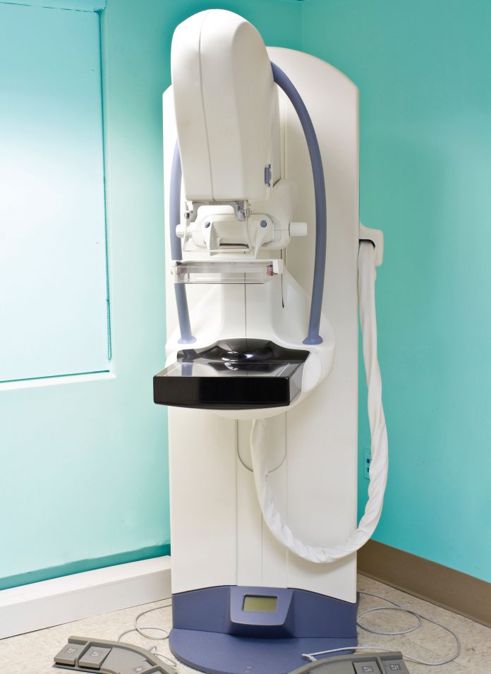
Automated Breast Ultrasound (ABUS)
Making Mammography Obsolete
Abstract There is a growing body of evidence that suggests the harms associated with standard breast cancer screening exams (mammography, breast self-examinations, and clinical breast exams) may outweigh the benefits. These harms include false-positive results, over-diagnosis, and over-treatment. Automated breast ultrasound (ABUS) is one of the latest technological breakthroughs that have been proposed as a suitable alternative for breast cancer screening. ABUS is a safe, painless, radiation-free, and non-invasive technology. It is a 3D ultrasound technology that is specifically developed for whole breast imaging and allows for images of high-resolution to be produced. Although somewhat limited, the currently available evidence is reviewed and appears to support the use of ABUS as a valuable screening tool, especially when considering the risks and benefits of the available alternatives. ABUS has also been studied in comparison to mammography, hand-held ultrasound, and MRI. In all studies, ABUS appears to provide additional value in terms of its sensitivity and specificity. ABUS appears to have high accuracy in determining preoperative cancer lesions and appears to be accurate in analyzing all types of histological subgroups. The main limitation in the studies that have been conducted is that they included a higher proportion of malignant lesions or breast masses than would be found in the screening population. Other limitations are that interpreting ABUS data requires a learning curve and that ABUS may be less valuable when it comes to examining the axillary regions or when examining the vascularity and elasticity of breast lesions. Largerscale research will need to be conducted on this exciting technology to determine its exact role in breast cancer screening, but ABUS will likely prove to be a cost-effective alternative.
In Canada, regular screening for breast cancer occurs with mammography, breast self-examinations, and clinical breast exams (Shields 2009). Currently, there exists controversy over exactly which screening tools should be utilized for particular patient populations. These three screening exams are recommended to reduce mortality due to breast cancer, but there is a growing body of evidence that suggests that the harms associated with these exams may outweigh the benefits (Canadian Task Force 2011). These harms include false-positive results, over-diagnosis, and over-treatment. In addition, positive results often cause emotional pain and anxiety for patients that can be severe and have long-term implications (Montgomery 2010). As such, there is a high demand for an alternative that reduces these harms and yet provides clinically useful information.
Automated breast ultrasound (ABUS) is one of the latest technological breakthroughs that have been proposed as a suitable alternative for breast cancer screening (Wenkel 2008). ABUS is a safe, painless, radiation-free, and non-invasive technology. It is a 3D ultrasound technology that is specifically developed for whole breast imaging and allows for images of high-resolution to be produced (Lin 2011). It has also been referred to in the literature as automated breast volume scanning (ABVS), automated whole-breast ultrasound (AWBS), and sonographic tomography (Kelly 2010). In this article, it will only be referred to as ABUS for simplicity. Although somewhat limited, the currently available evidence appears to support the use of ABUS as a valuable screening tool, especially when considering the risks and benefits of the available alternatives. This article will review the studies that have been conducted and discuss the value of ABUS compared to other breast imaging methods.
A recent pilot study of ABUS was conducted to evaluate its ability to detect and classify breast lesions according to the Breast Imaging Reporting and Data System (BI-RADS) (Wenkel 2008). Thirty-five women who had unclear findings in breast diagnosis performed through palpation, sonography, or mammography were selected and received ABUS. Five radiologists independently evaluated the ABUS images and classified them according to BI-RADS. All breast cancers were found with the ABUS by all examiners and correctly classified in the BI-RADS. In this study, ABUS allowed detection of solid and cystic lesions and their BI-RADS classification with a high reliability. This study was not without limitations. The first limitation was that the selected patient group had a high number of lesions and thus did not reflect a screening population. Another limitation was that some histological subgroups such as medullary carcinoma and ductal carcinoma in situ were not present in the cohort and thus the researchers could not comment on the ability of ABUS to detect these subgroups. Finally, only the affected breast was scanned and analyzed, possibly resulting in a high degree of alertness by the radiologists.
Another recent study of ABUS was conducted to determine the accuracy of measuring preoperative cancer extent (Tozaki 2010). This retrospective study looked at forty patients with histopathologically confirmed breast cancer who underwent ABUS on the day prior to surgery. The age range of patients was 31-76 years old. The discrepancy of the tumor extent between ABUS and the histological examination was calculated. It was found that ABUS enabled visualization of the breast carcinomas in all patients. The histopathological diagnosis was ductal carcinoma in situ in seven patients and invasive ductal carcinoma in thirty-three patients. The accuracy of determination of the tumor extent with a deviation in length of less than 2cm was 98%. This particular study helps fill gaps in the evidence to support the use of ABUS for these particular histological subgroups.
The same researchers have also studied the optimal scanning technique to cover the whole breast using ABUS (Tozaki 2010). In this study, forty patients aged 23-68 years old underwent ABUS in the upper-outer, lower-outer, lower-inner, and upperinner breast regions. Three examiners were used. The researchers also compared ABUS to hand-held ultrasound (HHUS). In total, sixty-one lesions were detected by HHUS and this number was consistent with those found by ABUS. No comment could be made when comparing the diagnostic accuracy of ABUS due to the small number of histologically confirmed lesions. The total scanning time for ABUS ranged from 10-12 minutes, which is fairly reasonable. All four scanning techniques for the major segments of the breast were found to be operator-independent and feasible for performing ABUS. Some of the disadvantages for ABUS that were identified by this study included the difficulty in examining the axillary regions, as well as analyzing the vascularity and elasticity of the breast lesions. Overall, this study indicates that ABUS is comparable to HHUS with regards to scanning technique, but with the additional benefit of being operator-independent.
Breast cancer detection using mammography plus ABUS compared to mammography alone has been studied (Kelly 2010). Twelve radiologists were blinded and provided with images of radiographically dense breasts. Half of the images were malignant. The radiologists first reviewed the mammograms and assessed their likelihood ratings based on BI-RADS and the Digital Mammographic Imaging Screenings Trial (DMIST). The radiologists then reviewed the mammograms with the ABUS data. Performance across screening techniques was compared using absolute callback, areas under the curve, and with figure of merit. With ABUS, true positives of cancer detection increased by 63% with only a 4% decrease in true negatives. The area under the curve average and the figure of merit were higher when mammography was paired with ABUS compared to mammography alone. Therefore, this study concluded that for dense-breasted women, incorporating ABUS with mammography adds significant clinical value. The automated process for breast ultrasound eliminates operator variability, provides greater consistency, and ensures reproducibility of quality images. Similar to one of the studies that were discussed previously, one limitation of this study might have been that the radiologists could have been extremely vigilant by realizing that the test images had higher rates of lesions than in a normal population.
ABUS has also been compared to MRI in a retrospective analysis (Grady 2010). Forty-one women who were diagnosed with breast cancer had preoperative staging using ABUS. They subsequently underwent bilateral contrast-enhanced MRI prior to surgery to determine tumor extension and other information regarding the spread of the cancer. Both imaging techniques were compared for accuracy to pathologic truth. ABUS accurately staged breast cancer preoperatively in 68% of cases, while MRI only did so in 54% of cases. ABUS resulted in an overall improvement in accuracy of 14% and thus appears superior to contrast-enhanced bilateral breast MRI. A second more recent study has also compared ABUS to MRI and determined how closely their results match (Moon 2011). ABUS and MRI breast images were obtained from forty patients; bilaterally in twenty-seven patients and unilaterally in thirteen patients. Calculated values for percent density and breast volume were compared and found to be highly correlated. These studies together appear to support ABUS as being equal to or superior to MRI.
The diagnostic accuracy of ABUS, including its sensitivity and specificity has been studied (Wojcinski 2011). In one study, ABUS data sets from fifty patients were collected and a database was created containing twenty-three women who had no detectable lesions with conventional ultrasound, thirteen women with benign lesions, and fourteen women with known breast cancer. An independent examiner evaluated the ABUS data on a separate workstation without any prior knowledge of the patients. The diagnostic accuracy for the ABUS was 66.0%. All breast cancers were detected, resulting in a sensitivity of 100%.
However, due to the high number of requests for second-look ultrasounds, specificity was 52.8%. In this study, the researchers concluded that ABUS must still be regarded as an experimental technique. The main limitations identified in this study were that the design had a limited sample size and that the proportion of cases to controls was not representative of the whole population.
The difference between ABUS and HHUS in detecting and diagnosing breast lesions has been examined and has provided further data on its diagnostic value (Lin 2011). In this study, eighty-one patients were subjected to both examinations and the number of lesions detected was compared. Diagnostic accuracy and specificity were calculated. Ninety-five lesions were detected by both devices. Both exhibited a sensitivity of 100% and a high specificity (ABUS 95.0% and HHUS 85.0%). ABUS had a higher diagnostic accuracy (97.1%) than HHUS (91.4%) for breast neoplasms. ABUS was also capable of displaying the retraction phenomenon in coronal plane, which produced a high specificity (100.0%) and a high sensitivity (80.0%) in detecting breast cancer, while it also had a high accuracy (91.4%) in determining malignant from benign lesions. A second study also looked at the differences between ABUS and HHUS (Isobe 2011). Sixty patients underwent ABUS and HHUS and four segments were scanned. In fourteen of the fifteen patients with breast lesions under the nipple, the lesions were detectable with both HHUS and ABUS. In the other patient, the lesion was not detectable by HHUS but was detected by ABUS. Overall, these two studies show that ABUS likely has superior diagnostic accuracy when compared to HHUS.
The inter-observer agreement of radiologists looking at breast masses that were detected by ABUS has been evaluated (Zhang 2011). In this study, 208 patients were subjected to ABUS and data were automatically sent to the ABUS workstation. Two radiologists evaluated 234 breast masses (148 benign and 86 malignant). The reviewers were blinded to the patient’s mammographic images, medical history, and pathologic findings. Substantial agreement was obtained for lesion shape, orientation, margin, echo pattern, posterior acoustic features, calcification, and final assessment. Fair agreement was obtained for retraction phenomenon and lesion boundary. This study provides evidence that inter-observer agreement for ABUS is high.
Researchers have retrospectively evaluated the detection of benign and malignant breast masses using ABUS data to determine which lesion variables affect detectability (Chang 2011). In this study, bilateral whole breast images were obtained using ABUS in sixtyseven consecutive women who were scheduled to undergo needle biopsy due to suspicious breast masses. Twenty-four invasive ductal cancers in twenty-three breasts, forty-six benign breast lesions in forty-four breasts, and thirty-eight normal breasts were included. Three breast radiologists who did not perform the examinations and were blinded to the histology reviewed the ABUS data. Sensitivities for benign mass detection ranged from 56.3% to 66.7% and for malignant masses ranged from 87.5% to 95.8%, indicating significantly higher sensitivity for malignant breast masses. The overall specificity was 79.5%. An analysis showed that mass size, surrounding tissue changes, and shape of the mass were the variables associated with detectability at ABUS. The lower detection rate for benign lesions was attributed to the fact that they only included solid breast lesions and not cystic lesions, which are easier to detect. This study was limited by the patient group that had a high number of lesions and did not represent a screening population. There also exists a learning curve in interpreting ABUS and these particular radiologists had little prior experience with ABUS, which may have affected the results.
In terms of financial cost, ABUS will probably prove cost-effective as a supplement to mammography for women with dense breasts (Feig 2010). Once radiologists have had practice with interpreting ABUS data and have progressed along the learning curve, it is likely that ABUS will provide clinicians with valuable diagnostic information. The ABUS system has an excellent safety profile and it is painless, radiation-free, and non-invasive. The evidence available demonstrates that it is specifically developed for whole breast imaging, but that it may be less valuable when it comes to examining the axillary regions or when examining the vascularity and elasticity of breast lesions. Some of the other variables that may limit its ability to detect breast lesions also include mass size and shape as well as surrounding tissue changes. Even so, ABUS appears to have high accuracy in determining preoperative cancer lesions and appears to be accurate in analyzing all types of histological subgroups. The evidence available seems to suggest that ABUS performs better than mammography, MRI, and HHUS and that inter-rater agreement for ABUS data are high. Its sensitivity and specificity are both high, especially when comparing these values to those of alternative screening methods. Many of the studies that have been done included a higher proportion of malignant lesions or breast masses than would be found in the screening population, meaning that it is likely too early to recommend ABUS as an independent screening tool. Larger-scale research will need to be conducted on this exciting technology to determine its exact role in breast cancer screening.
References
Canadian Task Force on Preventive Health Care, Tonelli M, Gorber SC, Joffres M, Dickinson J, Singh H, Lewin G, Birtwhistle R, Fitzpatrick-Lewis D, Hodgson N, Ciliska D, Gauld M, Liu YY. Recommendations on screening for breast cancer in average-risk women aged 40-74 years. CMAJ. 2011;183(17):1991-2001.
Chang JM, Moon WK, Cho N, Park JS, Kim SJ. Radiologists’ performance in the detection of benign and malignant masses with 3D automated breast ultrasound (ABUS). Eur J Radiol. 2011;78(1):99-103.
Feig S. Cost-effectiveness of mammography, MRI, and ultrasonography for breast cancer screening. Radiol Clin North Am. 2010;48(5):879-91.
Grady I, Gorsuch-Rafferty H, Hansen P. Sonographic tomography for the preoperative staging of breast cancer prior to surgery. Journal of Ultrasound. 2010;13(2):41-45.
Isobe S, Tozaki M, Yamaguchi M, Ogawa Y, Homma K, Satomi R, Saito M, Joo C, Fukuma E. Detectability of breast lesions under the nipple using an automated breast volume scanner: comparison with handheld ultrasonography. Jpn J Radiol. 2011;29(5):361-5.
Kelly KM, Dean J, Lee SJ, Comulada WS. Breast cancer detection: radiologists’ performance using mammography with and without automated whole-breast ultrasound. Eur Radiol. 2010;20(11):2557-64.
Lin X, Wang J, Han F, Fu J, Li A. Analysis of eighty-one cases with breast lesions using automated breast volume scanner and comparison with handheld ultrasound. Eur J Radiol. 2011. doi:10.1016/j.ejrad.2011.02.038
Montgomery M and McCrone SH. Psychological distress associated with the diagnostic phase for suspected breast cancer: systematic review. Journal of Advanced Nursing. 2010; 66(11), 2372–2390.
Moon WK, Shen YW, Huang CS, Luo SC, Kuzucan A, Chen JH, Chang RF. Comparative study of density analysis using automated whole breast ultrasound and MRI. Med Phys. 2011;38(1):382-9.
Shields M, Wilkins K. An update on mammography use in Canada. Health Rep. 2009;20(3):7-19.
Tozaki M, Fukuma E. Accuracy of determining preoperative cancer extent measured by automated breast ultrasonography. Jpn J Radiol. 2010;28(10):771-3.
Tozaki M, Isobe S, Yamaquchi M, Ogawa Y, Kohara M, Joo C, Fukama E. Optimal scanning technique to cover the whole breast using an automated breast volume scanner. Jpn J Radiol. 2010;28(4):325-8.
Wenkel E, Heckmann M, Heinrich M, Schwab SA, Uder M, Schulz-Wendtland R, Bautz WA, Janka R. Automated breast ultrasound: lesion detection and BI-RADS classification—a pilot study. Rofo. 2008;180(9):804-8.
Wojcinski S, Farrokh A, Hille U, Wiskirchen J, Gyapong S, Soliman AA, Degenhardt F, Hillemanns P. The Automated Breast Volume Scanner (ABVS): initial experiences in lesion detection compared with conventional handheld B-mode ultrasound: a pilot study of 50 cases. Int J Womens Health. 2011;3:337-46.
Zhang J, Lai XJ, Zhu QL, Wang HY, Jiang YX, Liu H, Dai Q, You SS, Xiao MS, Sun Q. Interobserver agreement for sonograms of breast lesions obtained by an automated breast volume scanner. Eur J Radiol. 2011; doi:10.1016/j.ejrad.2011.06.043


